Download
1 / 37
400 likes | 1.14k Views
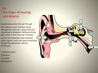
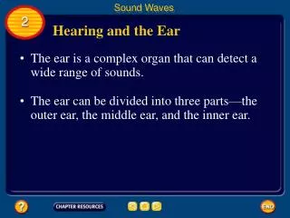
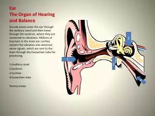
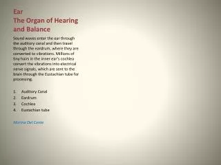
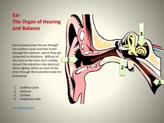
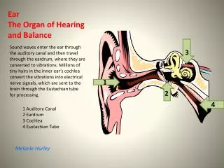
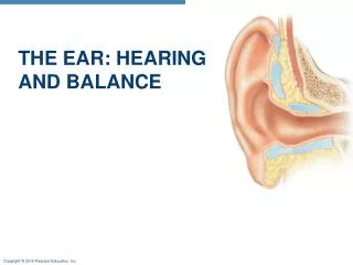
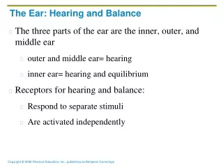
The Ear: Hearing and Balance. The three parts of the ear are the inner, outer, and middle ear The outer and middle ear are involved with hearing The inner ear functions in both hearing and equilibrium Receptors for hearing and balance: Respond to separate stimuli Are activated independently.

E N D
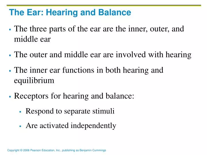
The Ear: Hearing and Balance • The three parts of the ear are the inner, outer, and middle ear • The outer and middle ear are involved with hearing • The inner ear functions in both hearing and equilibrium • Receptors for hearing and balance: • Respond to separate stimuli • Are activated independently
The Ear: Hearing and Balance Figure 15.25a
Middle and Internal Ear Figure 15.25b
Ear Ossicles Figure 15.26
Inner Ear Figure 15.27
Resonance of the Basilar Membrane Figure 15.32
The Cochlea Figure 15.28
Excitation of Hair Cells in the Organ of Corti Figure 15.28c
The Cochlea • A spiral, conical, bony chamber that: • Extends from the anterior vestibule • Coils around a bony pillar called the modiolus • Contains the cochlear duct, which ends at the cochlear apex • Contains the organ of Corti (hearing receptor)
The Cochlea • The cochlea is divided into three chambers: • Scala vestibuli • Scala media • Scala tympani
The Cochlea • The scala tympani terminates at the round window • The scalas tympani and vestibuli: • Are filled with perilymph • Are continuous with each other via the helicotrema • The scala media is filled with endolymph
The Cochlea • The “floor” of the cochlear duct is composed of: • The bony spiral lamina • The basilar membrane, which supports the organ of Corti • The cochlear branch of nerve VIII runs from the organ of Corti to the brain
Sound and Mechanisms of Hearing • Sound vibrations beat against the eardrum • The eardrum pushes against the ossicles, which presses fluid in the inner ear against the oval and round windows • This movement sets up shearing forces that pull on hair cells • Moving hair cells stimulates the cochlear nerve that sends impulses to the brain
Properties of Sound • Sound is: • A pressure disturbance (alternating areas of high and low pressure) originating from a vibrating object • Composed of areas of rarefaction and compression • Represented by a sine wave in wavelength, frequency, and amplitude
Properties of Sound • Frequency – the number of waves that pass a given point in a given time • Pitch – perception of different frequencies (we hear from 20–20,000 Hz)
Properties of Sound • Amplitude – intensity of a sound measured in decibels (dB) • Loudness – subjective interpretation of sound intensity Figure 15.29
Transmission of Sound to the Inner Ear • The route of sound to the inner ear follows this pathway: • Outer ear – pinna, auditory canal, eardrum • Middle ear – malleus, incus, and stapes to the oval window • Inner ear – scalas vestibuli and tympani to the cochlear duct • Stimulation of the organ of Corti • Generation of impulses in the cochlear nerve
Frequency and Amplitude Figure 15.30
Transmission of Sound to the Inner Ear Figure 15.31
Resonance of the Basilar Membrane • Sound waves of low frequency (inaudible): • Travel around the helicotrema • Do not excite hair cells • Audible sound waves: • Penetrate through the cochlear duct • Vibrate the basilar membrane • Excite specific hair cells according to frequency of the sound
The Organ of Corti • Is composed of supporting cells and outer and inner hair cells • Afferent fibers of the cochlear nerve attach to the base of hair cells • The stereocilia (hairs): • Protrude into the endolymph • Touch the tectorial membrane
Excitation of Hair Cells in the Organ of Corti • Bending cilia: • Opens mechanically gated ion channels • Causes a graded potential and the release of a neurotransmitter (probably glutamate) • The neurotransmitter causes cochlear fibers to transmit impulses to the brain, where sound is perceived
Auditory Pathway to the Brain • Impulses from the cochlea pass via the spiral ganglion to the cochlear nuclei • From there, impulses are sent to the: • Superior olivary nucleus • Inferior colliculus (auditory reflex center) • From there, impulses pass to the auditory cortex • Auditory pathways decussate so that both cortices receive input from both ears
Auditory Processing • Pitch is perceived by: • The primary auditory cortex • Cochlear nuclei • Loudness is perceived by: • Varying thresholds of cochlear cells • The number of cells stimulated • Localization is perceived by superior olivary nuclei that determine sound
Deafness • Conduction deafness – something hampers sound conduction to the fluids of the inner ear (e.g., impacted earwax, perforated eardrum, osteosclerosis of the ossicles) • Sensorineural deafness – results from damage to the neural structures at any point from the cochlear hair cells to the auditory cortical cells
Deafness • Tinnitus – ringing or clicking sound in the ears in the absence of auditory stimuli • Meniere’s syndrome – labyrinth disorder that affects the cochlea and the semicircular canals, causing vertigo, nausea, and vomiting
Mechanisms of Equilibrium and Orientation • Vestibular apparatus – equilibrium receptors in the semicircular canals and vestibule • Maintains our orientation and balance in space • Vestibular receptors monitor static equilibrium • Semicircular canal receptors monitor dynamic equilibrium
Anatomy of Maculae • Maculae are the sensory receptors for static equilibrium • Contain supporting cells and hair cells • Each hair cell has stereocilia and kinocilium embedded in the otolithic membrane • Otolithic membrane – jellylike mass studded with tiny CaCO3 stones called otoliths • Utricular hairs respond to horizontal movement • Saccular hairs respond to vertical movement
Anatomy of Maculae Figure 15.35
Inner Ear • Bony labyrinth • Tortuous channels worming their way through the temporal bone • Contains the vestibule, the cochlea, and the semicircular canals • Filled with perilymph • Membranous labyrinth • Series of membranous sacs within the bony labyrinth • Filled with a potassium-rich fluid
The Vestibule • The central egg-shaped cavity of the bony labyrinth • Suspended in its perilymph are two sacs: the saccule and utricle • The saccule extends into the cochlea
The Vestibule • The utricle extends into the semicircular canals • These sacs: • House equilibrium receptors called maculae • Respond to gravity and changes in the position of the head
The Vestibule Figure 15.27
The Semicircular Canals • Three canals that each define two-thirds of a circle and lie in the three planes of space • Membranous semicircular ducts line each canal and communicate with the utricle • The ampulla is the swollen end of each canal and it houses equilibrium receptors in a region called the crista ampullaris • These receptors respond to angular movements of the head
The Semicircular Canals Figure 15.27