Download
1 / 62
650 likes | 944 Views
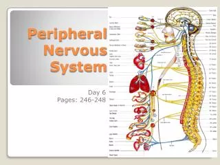
PERIPHERAL NERVOUS SYSTEM REVIEW (Spinal Nerve Review). Spinal Nerves. Dorsal root – contains sensory fibers Cell bodies – located in the dorsal root ganglion Ventral root – contains motor fibers arising from anterior gray column. Branch into dorsal ramus and ventral ramus

E N D
Spinal Nerves Dorsal root – contains sensory fibers • Cell bodies – located in the dorsal root ganglion Ventral root – contains motor fibers arising from anterior gray column • Branch into dorsal ramus and ventral ramus • Dorsal and ventral rami contain sensory and motor fibers • Rami communicantes connect to the base of the ventral ramus • Lead to the sympathetic chain ganglia Figure 14.7a
Innervation of the Back DORSAL ROOT DORSAL ROOT GANGLION VENTRAL ROOT SPINAL NERVE DORSAL RAMI VENTRAL RAMI RAMI COMMUNICANTES SYMPATHETIC TRUNK GANGLION A B C D E H F G Figure 14.7b
Introduction to Nerve Plexuses • Nerve plexus – network of nerves • Ventral rami • Branch and join with one another • Form nerve plexuses • In cervical, brachial, lumbar, and sacral regions • Primarily serve the limbs Which spinal nerves do not contribute to Nerve plexuses? T2 – T12
The Cervical Plexus • Buried deep in the neck • Under the sternocleidomastoid muscle • Formed by ventral rami of first four cervical nerves ( C1 – 4) • Some innervate muscles of the anterior neck • Phrenic nerve – the most important nerve of the cervical plexus ? ?
The Cervical Plexus • Buried deep in the neck • Under the sternocleidomastoid muscle • Formed by ventral rami of first four cervical nerves ( C1 – 4) • Some innervate muscles of the anterior neck • Phrenic nerve – the most important nerve of the cervical plexus
The Brachial Plexus and Innervation of the Upper Limb • Brachial plexus lies in the neck and axilla • Formed by ventral rami of C5 – C8 Figure 14.9d
The Brachial Plexus and Innervation of the Upper Limb • What Cervical Root gives rise to the middle trunk of the brachial plexus? Figure 14.9d
The Brachial Plexus and Innervation of the Upper Limb • What Cervical Root gives rise to the middle trunk of the brachial plexus? Figure 14.9d
The Brachial Plexus and Innervation of the Upper Limb What Cord from the Brachial plexus gives rise to the Median nerve? Figure 14.9d
The Brachial Plexus and Innervation of the Upper Limb What Cord from the Brachial plexus gives rise to the Median nerve? Figure 14.9d
The Brachial Plexus and Innervation of the Upper Limb This nerve is a continuation of the posterior cord? Figure 14.9d
The Brachial Plexus and Innervation of the Upper Limb This nerve is a continuation of the posterior cord? Figure 14.9d
The Brachial Plexus and Innervation of the Upper Limb The Ulnar nerve branches from what cord? Figure 14.9d
The Brachial Plexus and Innervation of the Upper Limb What is the main branch of theLateral cordfrom the Brachial plexus? Figure 14.9d
The Brachial Plexus Figure 14.9a
Posterior Cord extension - Axillary and Radial Nerves Figure 14.11
The Lumbar Plexus and Innervation of the Lower Limb • Lumbar plexus • Arises from L1– L4 • Smaller branches innervate the posterior abdominal wall and psoas muscle • Main branches innervate the anterior thigh
The Lumbar Plexus Figure 14.12a, b
The Sacral Plexus • Arises from spinal nerves L4–S4 • Caudal to the lumbar plexus • Often considered with the lumbar plexus • Lumbosacral plexus
Innervation of the Lower Limb • Sciatic nerve – the largest nerve of the sacral plexus • Actually two nerves in one sheath • Tibial nerve – innervates most of the posterior lower limb • Common fibular (peroneal) nerve – innervates muscles of the anterolateral leg ? ?
Innervation of the Lower Limb • Sciatic nerve – the largest nerve of the sacral plexus • Actually two nerves in one sheath • Tibial nerve – innervates most of the posterior lower limb • Common fibular (peroneal) nerve – innervates muscles of the anterolateral leg
Innervation of the Lower Limb • Superior and inferior gluteal nerves • Innervate the gluteal muscles • Pudendal nerve • Innervates muscles of the perineum
The Sacral Plexus ? ? Figure 14.13
The Sacral Plexus Figure 14.13
Innervation of the Skin: Dermatomes • Dermatome – an area of skin • Innervated by cutaneous branches of a single spinal nerve • Upper limb • Skin is supplied by nerves of the brachial plexus • Lower limb • Lumbar nerves – anterior surface • Sacral nerves – posterior surface
Map of Dermatomes – Anterior View Figure 14.14a
Map of Dermatomes – Posterior View Figure 14.14b
Map of Dermatomes – Anterior View Which spinal nerve cutaneous branch is damaged when a person loses sensation at the Digits 1 and 2 (right hand)? Figure 14.14a
Map of Dermatomes – Anterior View Figure 14.14a
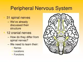
Cranial Nerves • Attach to the brain and pass through foramina of the skull • Numbered from I–XII • Which Cranial nerves are attached to the forebrain? • All others attach to the brain stem • Which Cranial nerves are attached to the Midbrain? • at the Pons? • at the Medulla? • Primarily serve head and neck structures • Which of Cranial nerve extends into the abdomen? CN I and II CN III and IV CN V and VII CN VIII and XII CN X
The 12 Pairs of Cranial Nerves CN I CN II CN III CN IV CN V CN VI CN VII CN VIII CN IX CN X CN XI CN XII K L A J B I C D H G F E Figure 14.5
Olfactory Nerves Limbic lobe Piriform lobe • Sensory nerves of smell Table 14.3 (1 of 12)
Optic Nerve • Sensory nerve of vision Table 14.3 (2 of 12)
Oculomotor Nerve (EOM) • Innervates four of the extrinsic eye muscles Table 14.3 (3 of 12)
Oculomotor Nerve (Pupillary Constriction) • Innervates four of the extrinsic eye muscles Pupillary constriction Table 14.3 (3 of 12)
Trochlear Nerve • Innervates the superior oblique muscle (an extrinsic eye muscle) Superior midbrain Table 14.3 (4 of 12)
Abducens Nerve • Abducts the eyeball – innervates lateral rectus muscle Inferior Table 14.3 (6 of 12)
Medial Rectus Superior Rectus Inferior Rectus Inferior Oblique (Levator Palpebrae) Superior Oblique EXTRAOCULAR MUSCLS Lateral Rectus CN III CN IV CN VI LASOT
Trigeminal Nerve • Provides sensory innervation to the face • Motor innervation to chewing muscles (THROUGH THE MANDIBULAR DIVISION)
Trigeminal Nerve OPTHALMIC Pons Trigeminal n. Trigeminal gang. Sup. Orbital fissure Supraorbital foramen MAXILLARY Pons Trigeminal n. Trigeminal gang. Foramen ovale Mandibular foramen Mental foramen foramen MAXILLARY Pons Trigeminal n. Trigeminal gang. Foramen rotundum Infraorbital foramen Table 14.3 (5 of 12)
Facial Nerve • Innervates muscles of facial expression • Pons • Internal Acoustic Meatus • Chorda tympani to taste • Anterior 2/3 tongue • 2) Somatic motor to facial • Muscles • 3) Parasympathetic to • Pterygopalatine (lacrimal) • and Submandibular (salivary) • ganglion - Table 14.3 (7 of 12)
Vestibulocochlear Nerve • Sensory nerve of hearing and balance Table 14.3 (8 of 12)
Glossopharyngeal Nerve • Innervates structures of the tongue and pharynx • Medulla • CN IX • Superior gang. • Inferior gang. • Jugular foramen • Parasymp. to parotid • gland via Otic gang. • 2) Carotid sinus • 3) Tongue • 4) Pharynx and throat Table 14.3 (9 of 12)
Vagus Nerve • A mixed sensory and motor nerve • “Wanders” into thorax and abdomen • Parasympathetic innervation of organs Medulla Jugular foramen Thorax and Abdomen Table 14.3 (10 of 12)
Accessory Nerve • An accessory part of the vagus nerve • Innervates trapezius muscle Cranial + Spinal root from medulla Jugular foramen 1) Cranial root goes w/ Vagus n. 2) Spinal root to a) Sternocleidomastoid b) Trapezius Table 14.3 (11 of 12)
Hypoglossal Nerve • Runs inferior to the tongue • Innervates the tongue muscles Medulla Hypoglossal canal Tongue (movement) Table 14.3 (12 of 12)
PERIPHERAL NERVOUS SYSTEM REVIEW (Autonomic Nervous System Review))
The Peripheral Nervous System • Autonomic nervous system (ANS) • General visceral motor part of the PNS • ANS has two divisions • Parasympathetic • Sympathetic