Download
1 / 6
210 likes | 1.71k Views
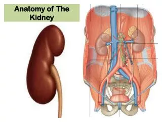
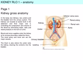
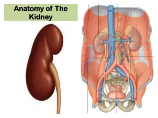
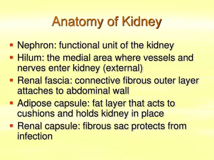
Anatomy of Kidney. Nephron: functional unit of the kidney Hilum: the medial area where vessels and nerves enter kidney (external) Renal fascia: connective fibrous outer layer attaches to abdominal wall Adipose capsule: fat layer that acts to cushions and holds kidney in place

E N D
Anatomy of Kidney • Nephron: functional unit of the kidney • Hilum: the medial area where vessels and nerves enter kidney (external) • Renal fascia: connective fibrous outer layer attaches to abdominal wall • Adipose capsule: fat layer that acts to cushions and holds kidney in place • Renal capsule: fibrous sac protects from infection
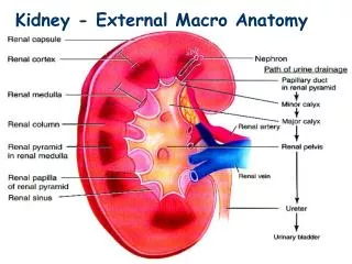
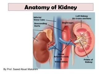
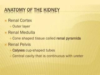
Anatomy of Kidney • Renal sinus: medial area where vessels and urine ducts enter kidney (internal) • Parenchyma: glandular tissue that forms urine • Outer renal cortex: thin outer layer of tissue that extends in columns dividing medulla into renal pyramids • Inner renal medulla: contains renal pyramids that funnel into the minor calyx
Anatomy of Kidney • Minor calyx: a small funnel like structure that collects urine from a renal pyramid • Major calyx: a larger funnel like structure that several minor calyx merge into • Renal Pelvis: an even larger funnel that several major calyx merge into • Glomerulus: a mass of capillaries that is enclosed by the nephron glomerular capsule (Bowman’s capsule)
Nephron • The nephron has two parts • Renal corpuscle: has two parts the glomerulus and the bowman’s capsule. • Renal tubule: a duct that leads away from the capsule and ends at the minor calyx. It is made up of several parts: PCT, nephron loop, DCT, collecting duct, papillary duct
Renal Tubule • Proximal convoluted tubule (PCT): contained in the renal cortex it follows the bowman’s capsule, is the longest part of the renal tubule, has microvilli to increase absorption. • Nephron loop: follows the PCT, has a descending thin segment and a ascending thick segment, active in salt transport, contained in the renal medulla
Renal Tubule • Distal convoluted tubule (DCT): when the nephron loop returns to the renal cortex, contains no microvilli • Collecting duct: a long straight tubule that several DCTs empty into • Papillary duct: a larger tubule that several collecting ducts empty into, the papillary duct enters the minor calyx