Download
1 / 18
250 likes | 449 Views
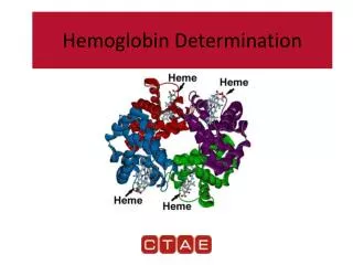
Introduction To Medical Technology. - Lab 16 -. Hemoglobin Concentration Determination. Hemoglobin (Hb). Hemoglobin (Hb) is the standard abbreviation for hemoglobin, the oxygen-carrying pigment and predominant protein in the red blood cells.

E N D
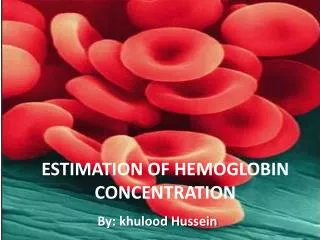
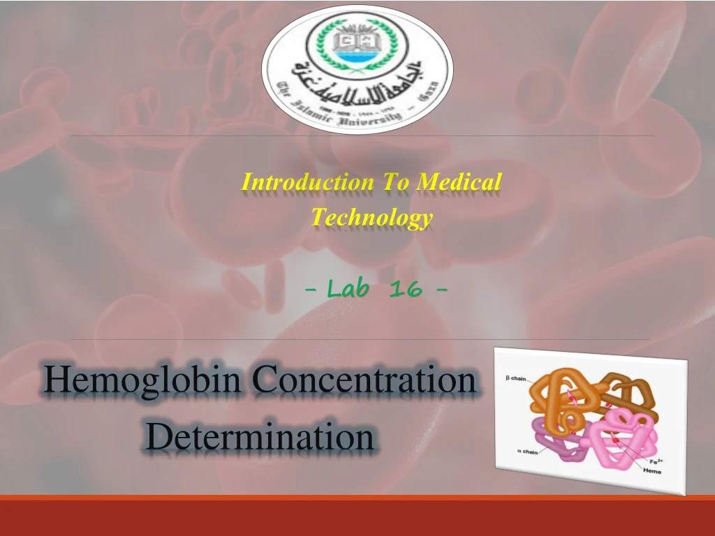
Introduction To Medical Technology - Lab 16 - Hemoglobin Concentration Determination
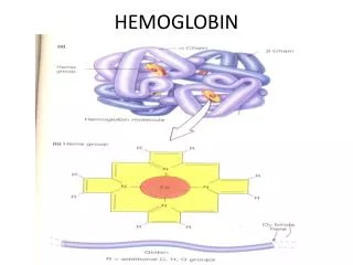
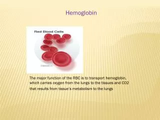
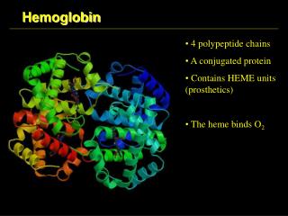
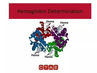
Hemoglobin (Hb) • Hemoglobin (Hb) is the standard abbreviation for hemoglobin, the oxygen-carrying pigment and predominant protein in the red blood cells. • Hemoglobin carries oxygen from places of high oxygen pressure (lungs) to places of low oxygen pressure (tissues), where it readily releases the oxygen. Hemoglobin also returns CO2 from the tissues to the lungs. • A hemoglobin molecule consists of 1 molecule of globin and 4 molecules of heme (each containing 1 molecule of iron in the ferrous state).
Structure of hemoglobin • “Globin" consists of four polypeptide chains: two alpha chains, each with 141 amino acids and two beta chains, each with 146 amino acids. • The α and β globin chains are very similar in structure and each one of them is linked with a heme molecule. Each heme group can combine with 1 molecule of oxygen or CO2. • A heme group is a flat ring molecule containing carbon, nitrogen and hydrogen atoms, with a single Fe2+ ion at the center. Without the iron, the ring is called a porphyrin.
Normal Ranges • In the very common laboratory tests for hemoglobin (Hb), it is measured as total hemoglobin and the result is expressed as the amount of hemoglobin in grams (gm) per deciliter (dl) of whole blood, a deciliter being 100 milliliters. • The Normal Ranges For Hemoglobin Depend On: • The age. • Altitude • The sex of the person. • Normal values in an adult are 12 to 18 grams per deciliter (100 milliliters) of blood.
Secondary polycythemia which is may be due to: Dehydration (sever burns, diarrhea, vomitting, …etc.). Severe lung or heart disease. Living at high altitudes. Heavy smoking. Primary polycythemia which is due malignant variation in blood cells production in bone marrow Above-normal hemoglobin levels is called polycythemia which is may be:
Iron deficiency or deficiencies in essential vitamins of other elements, such as B12, folate, B6. Inherited hemoglobin defects, such as sickle cell anemia or Thalassemia. Other inherited defects affecting the red blood cells. Excessive bleeding. Excessive destruction of red blood cells. Kidney disease. Bone marrow failure or aplastic anemia. Cancers that affect the bone marrow. Below-normal hemoglobin levels may lead to anemia that can be the result of:
Measurement of hemoglobin • Principle: • Whole blood is diluted in a solution of potassium Ferricyanide and potassium cyanide. • The Hb is oxidized to met-hemoglobin by the potassium Ferricyanide. • The potassium cyanide then converts the met-hemoglobin to cyanmet-hemoglobin. The Cyan-methemoglobin Method for Hb determination is the reference method. • Hb (Fe++) K3Fe (CN)6Methemoglobin (Fe+++ ) KCN • Cyan-Methemoglobin
The absorbance of the cyanmet-hemoglobin at 540 nm is directly proportional to the Hb concentration. • Sulf-hemoglobin is not converted to cyanmet-hemoglobin; therefore, it can not be measured by this method.
Procedure of standard curve • Standard Curve Preparation • Create a standard curve, using a commercially available cyan-methemoglobin standard which, has constant concentration 25g/dl, the following dilutions should be made to get the line between the concentration & the absorbance of the standard using also drabkin reagent as shown:
Procedure • Allow the tubes to stand for 10 minutes. • Transfer the dilutions to cuvettes. Starting with the blank, zeroing the spectrophotometer with the BLANK solution, then measure the absorbance on a spectrophotometer at 540 nm. • Plot absorbance on the y-axis and the Hb concentration on the x-axis. The Hb concentrations of the patients’ samples and controls can be read from this standard curve.
Patient Sample Preparation • Pipette 5 ml of Cyan-methemoglobin reagent into a tube. Add 20 l of the sample into the tube. • Allow the tube to stand for 10 minutes. • Read Absorbance (A) in the spectrophotometer at 540 nm, zeroing the spectrophotometer with the BLANK solution.
Standard Curve calculation ∆ Y ∆ X Concentration of Standard
Discussion • mechanical sources of error: • Pipetting error. • Use of dirty or scratched cuvettes. • Use of deteriorated reagents. • Before the test sample is read, the solution should be clear: • A high WBC count: centrifuge specimen and use the supernatant for reading. • Hemoglobin S (HbS) and Hemoglobin C (HbC), dilute the mixture 1:1 with distilled water and then read in the colorimeter; multiply the reading by 2. • Lipemiacan also interfere, and a false result can be corrected by adding 0.02 ml of the patient’s plasma to 5 ml of the cyanmethemoglobin reagent, this solution being used as the reagent blank.
Drabkin’s reagent is sensitive to light. It should be stored in a brown bottle or in dark place. • Carboxy-hemoglobin takes up to 1 hr to convert to cyan-methemoglobin and therefore, theoretically could cause erroneous results in the samples from heavy smokers. However the degree of error is probably not clinically significant. • Because Drabkin’s reagent contains cyanide, it must be used cautiously; a minimum of four L of reagent is lethal. • Acid free sinks should be used for disposal of reagent and samples, because acidification of cyanide releases hydrogen cyanide gas. Copious amounts of water should be used to flush the sink after disposable.