Download
1 / 65
810 likes | 2.41k Views
AIRWAY MANAGEMENT – establishing, maintaining & removing artificial airway with complications. Dr. Poonam Patel. AIRWAY MANAGEMENT. Assessment – Mallampati score, mouth opening, thyromental distance Securing & maintenance – airway devices Artificial airway Supraglottic airway devices

E N D
AIRWAY MANAGEMENT – establishing, maintaining & removing artificial airway with complications. Dr. Poonam Patel
AIRWAY MANAGEMENT • Assessment – Mallampati score, mouth opening, thyromental distance • Securing & maintenance – airway devices • Artificial airway • Supraglottic airway devices • Tracheal tube • Devices for difficult airway • Management of complications
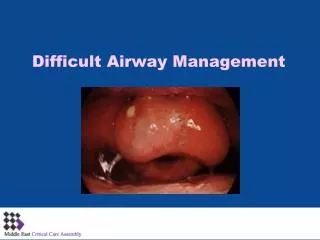
AIRWAY ASSESSMENT • Cervical spine movement • T-M joint movement • Mouth opening • Modified Mallampati grading • Thyromental distance
ARTIFICIAL AIRWAY Purpose of an airway – lift the tongue and epiglottis away from the posterior pharyngeal wall. Advantage of an airway – • Cervical spine movement does not occur when airway is inserted. • Decreased work of breathing during spontaneous respiration using a face mask. • Types Oropharyngeal airway Nasopharyngeal airway
AIRWAY ANATOMY • Normal • Obstructed airway
OROPHARYNGEAL AIRWAY Guedel airway – • Parts – flange, bite portion, air channel
OROPHARYNGEAL AIRWAY (contd.) • Sizes available • Colour coding
OROPHARYNGEAL AIRWAYS (contd.) • Uses – • To maintain open airway • Prevent endotracheal tube occlusion • Prevent tongue bite • Facilitate suction • Conduit for passing devices into oropharynx • Obtain a better mask fit • Contraindications – • Intact gag reflex • Oropharyngeal growth
OROPHARYNGEAL AIRWAY (contd.) • Pre requisite for insertion • Size estimation • Methods of insertion • Disadvantages - • Due to incorrect size • Laryngospasm in awake patient • Advantages - • 1) Simple to use, cheap. • 2) Not associated with sore throat • 3) Does not cause bacteremia
NASOPHARYNGEAL AIRWAY • Parts – flange, airway channel, bevel. • Size - inside diameter in millimeters. • Size determination • Method of insertion • Contraindications – 1) Anticoagulation 2) Basilar skull fracture 3) Nasal pathology, sepsis, or deformity of the nose or nasopharynx 4) History of epistaxis requiring medical treatment.
NASOPHARYNGEAL AIRWAY (contd.) • Uses of nasopharyngeal airway – • To maintain airway in patients with intact gag reflex • To facilitate suctioning • As a guide for a fiberscope or nasogastric tube • To apply continuous positive airway pressure (CPAP) • To dilate the nasal passages in preparation for nasotracheal intubation • To maintain the airway and administer anesthesia during dental surgery. • To maintain ventilation during oral fiberoptic endoscopy.
NASOPHARYNGEAL AIRWAY (contd.) • Advantages- 1) Nasal airway is better tolerated than an oral airway if the patient has intact airway reflexes. 2) Loose or poor dentition. 3) Trauma or pathology of the oral cavity. 4) It can be used when the mouth cannot be opened.
COMPLICATIONS OF ARTIFICIAL AIRWAY • Airway Obstruction • Trauma • Tissue Edema • Ulceration and Necrosis • Central Nervous System Trauma • Dental Damage • Laryngospasm and Coughing • Retention, Aspiration, or Swallowing • Devices Caught in Airway • Equipment Failure • Latex Allergy • Gastric Distention
SUPRAGLOTTIC AIRWAY DEVICES • Supraglottic devices fill a niche between the face mask and tracheal tube in terms of both anatomical position and degree of invasiveness. • These devices sit outside the trachea but provide a handsfree means of achieving a gas-tight airway.
SUPRAGLOTTIC AIRWAY DEVICES • Laryngeal Mask Airway Family – • LMA Classic • LMA Unique • LMA Flexible • LMA Fastrach • LMA CTrach • LMA Proseal 2) Other supraglottic airways similar to laryngeal mask – • Soft seal laryngeal mask • Ambu laryngeal mask • Intubating laryngeal airway 3) Other supraglottic airway devices • Laryngeal tube airway • Perilaryngeal airway • Streamlined pharynx airway liner
LARYNGEAL MASK AIRWAY FAMILY • LMA-Classic (standard LMA, Classic LMA, LMA-C, cLMA) • PARTS – • Curved tube (shaft) • Elliptical spoon-shaped mask • Two flexible vertical bars. • An inflatable cuff. • An inflation tube • Self-sealing pilot balloon. • 15-mm connector .
LMA CLASSIC • Insertion methods • Standard Technique • 180-degree Technique • Partial Inflation Technique • Thumb Insertion Technique
LMA-UNIQUE • Disposable laryngeal mask airway, DLMA). • It is made of polyvinylchloride • The dimensions are identical to the standard LMA, the tube is stiffer and the cuff less compliant. • Sizes • It may be a better choice for out-of-hospital or ward use. • Insertion and placement of the LMA-Unique is similar to the LMA-Classic. • The intracuff pressure increases significantly less in the LMA-Unique when nitrous oxide is used.
LMA-FLEXIBLE • The LMA-Flexible (wire-reinforced, reinforced LMA, RLMA, FLMA, flexible LMA) has a flexible, wire-reinforced tube. • The tube is longer and narrower. • Not available in sizes 1 and 1.5 • Useful for head and neck surgeries • Insertion method • Disadvantages - 1) The wire reinforcement does not prevent obstruction from biting. 2) The spiral reinforcing wire may become disrupted. 3) Only small sizes of tracheal tube or bronchoscope can pass through it. 4) Not preferred prolonged spontaneous ventilation. 5) Unsuitable for MRI scanning 6) Malposition is less easily diagnosed.
LMA-FASTRACH • The LMA-Fastrach (intubating LMA, ILMA, ILM, intubating laryngeal mask airway) – designed for tracheal intubation. • Parts – 1) A short, curved stainless steel shaft with a standard 15-mm connector. 2) Single, movable epiglottic elevator bar 3) A V-shaped guiding ramp built into the floor of the mask.
LMA-FASTRACH • Insertion technique • Uses • To facilitate tracheal intubation • It can also be used as a primary airway device. • Tracheal Intubation using LMA Fastrach – • Blind, • Blind nasal • Fiberscopic guided • Light guided
LMA-FASTRACH • Disadvantages • Pharyngeal pathology or limited mouth opening may make intubation difficult. • Cannot be used for intubation in patients below 30 kg. • The LMA-Fastrach tracheal tube is expensive & prolonged use is to be avoided. • The tracheal tube may be displaced downward or dislodged. • It should not be used in the prone position • Unsuitable for use in the MRI unit. • Increased incidence of sore throat and difficulty swallowing . • In patients with immobilized cervical spine, exerts pressure on the cervical spine.
LMA-CTrach • It has two built-in fiberoptic channels with a monitor. • Sizes - 3, 4, and 5 • Insertion technique • Advantages – • High first intubation attempt success rate with minimal neck movement. 2) Can be used during awake intubation in the presence of an unstable cervical spine. • Disadvantages 1) Poor image quality 2) The view may be obstructed by secretions, lubricant, or blood. 3) Cannot be used easily in the patient with a limited mouth opening.
LMA-ProSeal • Parts - cuff, inflation line with pilot balloon, airway tube, and drain (gastric access) tube. • Function of second dorsal cuff • Insertion techniques – introducer, guided, digital methods • Confirmation of proper placement
LMA-ProSeal • Uses • Can be used for both spontaneous and controlled ventilation. • Preferred in situations where higher airway pressures are required, better airway protection is desirable, and for surgical procedures in which intraoperative gastric drainage or decompression is needed • Useful in cases where it is important to avoid airway trauma. • Safe for use in an MRI unit
LMA-ProSeal Disadvantages - 1) The LMA-ProSeal is less suitable as an intubation device. 2) Higher resistance in spontaneously breathing patients than other devices. 3) Requires a greater depth of anesthesia for insertion. 4) Airway obstruction after insertion. 5) Gastric insufflation 6) The LMA-ProSeal has a shorter life span.
LARYNGEAL TUBE AIRWAY • Parts – • The airway tube is wide, curved, color coded on the connector. • single lumen that is closed at the tip. • Small (esophageal, distal) cuff near the blind distal tip • Large (oropharyngeal, pharyngeal) cuff near the middle of the tube
LARYNGEAL TUBE AIRWAY (Cont.) • 5) One inflation tube to inflate both light blue cuffs. • 6) Two anterior-facing, oval-shaped openings (ventilation holes) • 7) Side holes lateral to the top of the distal opening. • 8) A ramp leads from the posterior wall toward the main ventilatory outlet • Reusable silicone or single-use versions made of polyvinylchloride.
LARYNGEAL TUBE AIRWAY (Cont.) • Insertion technique • Advantages - 1) The LT is relatively easy to insert 2) It is well tolerated during emergence 3) Because the distal cuff fits over the esophageal inlet, the risk of gastric inflation is low 4) Can be used with both spontaneous and controlled ventilation 5) High ventilation pressures can be used.
Laryngeal Tube Airway (Cont.) 6) This device may be especially useful for resuscitation (“cannot intubate, cannot ventilate” situation , obstetrics after failed intubation, edentulous patients). 7) The incidence of complications such as sore throat, mouth pain, or dysphagia is low. 8) Regurgitated liquid is less likely to be aspirated with the LT • Disadvantage • Failure to ventilate if tube enters trachea – contrast combitube
ENDOTRACHEAL TUBE • The tracheal tube (endotracheal tube, intratracheal tube, tracheal catheter) is a device that is inserted through the larynx into the trachea to convey gases and vapors to and from the lungs. • Parts – • The machine (proximal) end • The patient (tracheal or distal) end • Bevel.
ENDOTRACHEAL TUBE 4) Murphy eye 5) A radiopaque marker 6) Cuff Systems - consists of the cuff plus an inflation system, which includes an inflation tube, a pilot balloon, and an inflation valve.
ENDOTRACHEAL TUBE Oral intubation – • Direct Laryngoscopy • Blind Oral Intubation • Digital Technique • Fiberoptic guided • Retrograde intubation Nasal intubation – • Direct Laryngoscopy • Flexible Fiberoptic Laryngoscopy • Blind Nasal Intubation
EXTUBATION • The tracheal tube (extubation) is removed when it is no longer needed for airway protection. • Extubation may be performed at different depths of anesthesia - “awake,” “light,” and “deep” • Preparation for Extubation • Initial Plan • Patient position plan • Bite block in place • Throat pack removed • Preoxygenation • Secretions aspirated from the pharynx (the trachea also if indicated)
EXTUBATION • Complications at Extubation • Hypoventilation (residual effect of anesthetic drugs and neuromuscular blockade) • Upper airway obstruction • Laryngospasm and bronchospasm • Coughing (wound disruption) • Impaired laryngeal competence and pulmonary aspiration • Hypertension, tachycardia, dysrhythmias, myocardial ischemia
FLEXIBLE FIBEROPTIC BRONCHOSCOPY • Indications – • Difficult intubation predicted • Congenital airway abnormalities • Acquired airway abnormalities • Trauma Contraindications- • Lack of time • Blood & secretions in oral cavity • Edema of pharynx or tongue • Technique – oral or nasal (awake or GA)
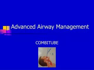
COMBITUBE • Device for difficult airway • PARTS – 1) Two separate lumens (pharyngeal & tracheoesophageal) that are fused longitudinally 2) Two inflatable cuffs. 3) Each lumen is linked by a short tube to a standard 15-mm connector at the breathing system end. 4) Pharyngeal lumen - occluded distal end and eight oval-shaped perforations (ventilating eyes) between the cuffs, coloured blue.
COMBITUBE 5) Tracheoesophageal lumen - patent distal end and a clear tube. 6) The smaller distal cuff serves to seal either the esophagus or trachea, depending on its placement. 7) The larger (pharyngeal) cuff (balloon) is above the perforations. 8) The pilot balloon for the pharyngeal cuff is colored blue.
COMBITUBE • Sizes: • Regular (41 [Fr]) • SA (37 Fr) • Recommended for patients with a height greater than 5 feet (152 cm). • Not recommended for patients younger than 12 years of age. • METHOD OF INSERTION
COMBITUBE • Indications • Airway management in the difficult-to-intubate patient • Massive airway bleeding or regurgitation. • Limited access to the airway and limited mouth opening • Cervical spine injury. • Useful in entertainers in whom it is important to avoid vocal cord damage. • In cardiopulmonary resuscitation in both prehospital and in-hospital settings. • “Cannot ventilate, cannot intubate” situation. • Can be used during percutaneous dilatational tracheostomy or tracheotomy
COMBITUBE • Contraindications • Active pharyngeal or laryngeal reflexes • Oesophageal trauma or pathology • ingestion of corrosive agents • Oropharyngeal, pharyngeal, or hypopharyngeal mass.
COMBITUBE • Advantages • Time needed for insertion is short and less skill is required • Can be inserted in presence of blood or secretions in the oropharynx. • Provides comparable ventilation and improved oxygenation to that of tracheal intubation • It can be used by an anesthesia provider having limited use of the left arm . • It is well tolerated by the patient during emergence from anesthesia. • Its use is not associated with high levels of trace gases. • Decreased risk of accidental extubation. • The Combitube provides good but not complete protection from aspiration
COMBITUBE • Disadvantages • Tracheal suctioning or fiberoptic bronchoscopy is not possible through the Combitube in the esophageal position • High airflow resistance • Insertion and removal of the Combitube is associated with a higher stress response • Trauma to the airway and esophagus • Sore throat and dysphagia. • Unsuitable for use in a patient with latex allergy . • The Combitube is expensive compared to other single use devices.
RETROGRADE INTUBATION • Retrograde (translaryngeal-guided, guided blind) intubation is an elective or emergency technique for securing a difficult airway, either alone or in conjunction with other techniques. • Retrograde intubation is a useful option in patients who cannot be intubated by using traditional techniques. • Procedure can be expected to take 5 minutes or more for completion.