Download
1 / 43
430 likes | 473 Views
Cerebellum. Coordination of movements. Vermis Hemispheres Folia, lobuli, lobi. Pars flocculonodularis. Fissura prima. Lobus ant. Lobus flocculonod. Lobus post. Fissura posterolat. Developmental anatomy. Archi- cerebellum. Afferents from vestib. labyrinth fish, amphibians.

E N D
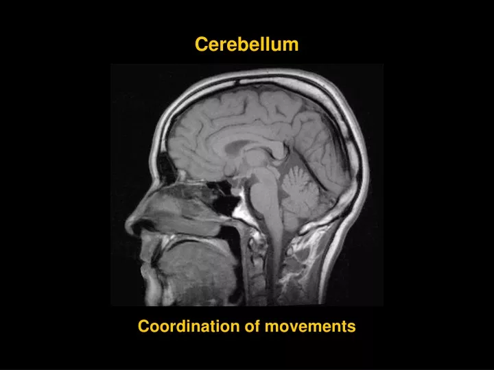
Cerebellum Coordination of movements
Vermis Hemispheres Folia, lobuli, lobi
Fissura prima Lobus ant. Lobus flocculonod. Lobus post. Fissura posterolat.
Developmental anatomy Archi- cerebellum Afferents from vestib. labyrinth fish, amphibians VESTIBULO -CEREBELLUM Afferents from spinal cord and brainstem reptiles, birds, mammals Paleo- cerebellum Paleo- cerebellum SPINO -CEREBELLUM Neo- cerebellum PONTO -CEREBELLUM Afferents from cortex telencephali
Structure of the cerebellum Grey matter Cortex cerebelli Nuclei cerebellares White matter Subst. medullaris laminae albae (arbor vitae) Pedunculi cerebellares
4 3 2 1 1ncl. dentatus2ncl. emboliformis3 ncll. globosi 4 ncl. fastigii Nuclei cerebelli
Spinocerebellum Ponto cerebellum 3 2 1 Vestibulocerebellum 1median zone 2paramedian zone 3 lateral zone L. flocculonodularis
Spinocerebellum Vestibulocerebellum Ponto cerebellum Vestibulocereb. ncll. vestibulares Spinocereb. ncll. fastigii, emboliformes, globosi Neocereb. ncl. dentatus
Pedunculi cerebel. inf. tr. sp-ce post., cuneo-ce, bulbo-ce, ve-ce, re-ce, olivo-ce from lobus flocculonodul. to ncll. vestibulares (tr. ce-ve), to RF of the brainstem (tr. ce-re) Pedunculi cerebel. medii tr. ponto-ce Pedunculi cerebel. sup. tr. sp-ce ant., ru-ce a afferents from ncl. mesenceph. CN V from ncll. emboliformes, globosi and dentatus Afferents : efferents = 40:1
Pathways of the cerebellum Afferents to the cortex cerebelli from vestib. labyrinth from spinal cord and brainstem from cortex of the brain Efferents from the nuclei to brainstem, thalamus
Function of the cerebellum ■ archicerebellum >posture and eye movements ■ paleocerebellum >progressive movements (walking, swimming etc.) ■ neocerebellum >manipulative movements and speech
CEREBELLAR DISORDERS Ataxia inability to stand upright without support Dysmetria„overshooting“- the hand may travel past the target Dyssynergiaincoordination Adiadochokinesia inability to perform rapid alternating movements
DIENCEPHALON ■thalamus (metathalamus) ■ epithalamus ■subthalamus ■hypothalamus
Thalamus ■tuberculum ant. ■pulvinar ■stria medullaris (tela choroidea ventr. III.) ■taenia choroidea (tela choroidea ventr. lat.) ■lamina affixa thalami ■stria terminalis (vena thalamostriata)
Metathalamus 1 2 1 corp. geniculatum med. brachium colliculi inf. – colliculus inf. 2 corp. geniculatum lat. brachium colliculi sup. – colliculus sup.
THALAMUS relay station of ascending pathways involved in motor circuits reciprocal connections to the association areas of the cerebral cortex – functions related to memory, cognition, judgement, mood
Anterior groupA ncll. ant. A DM LD LP VA Lateral groupdorsal row VL CM P VP: VPM LD ncl. lat. dors. LP ncl. lat. post. VPL R CGM CGL ventral row Medial groupDM ncl. dorsomed. Posterior groupP ncll. pulvinari,post. Intralaminar groupCM ncl. centromed. R ncll. reticulares VA ncl. ventr. ant. VL ncl. ventr. lat. VP ncl. ventr. post.: VPL ncl. ventr. post-lat VPM ncl. ventr. post-med CGL ncl. corporis gen. lat. CGM ncl. corporis gen. med.
Functional groups of nuclei ■ specific nuclei somatosensory sensory motor ■non-specific nuclei ■association nuclei
Specific nuclei VA VL VPL VPM GP CGM CGL auditorypathway cerebellum BG visual pathway RO SS:VPL, VPM S:CGM, CGL M:VA, VL tr. trig-th tr. so-th (taste) tr. sp-th LM
Non-specific nuclei ncll. intralaminares ncl. medianus R from FR of the brainstem and other thalamic nuclei to BG, thalamus, cortex (ARAS)
Projection to the cortex through specific and non-specific thalamic nuclei CORTEX THALAMUS specific pathway non-specific pathway
Association nuclei A DM LD LP P ■ integration of GSA a SA inputs to cortex ■reciprocal connections with the association cortex
Function of association nuclei cortex cortex Ncl. ant. thalami Interconnection of association areas of the cortex
Epithalamus ■ stria medullaris thalami ■trigonum habenulae ■commissura habenularum et post. ■corpus pineale (epiphysis cerebri)
Subthalamus ■ zona incerta ■ ncl. subthalamicus ■ part of subst. nigra Involved in motor circuits
Hypothalamus Corp. mamillaria Tuber cinereum Infundibulum Eminentia mediana Hypophysis cerebri Chiasma opticum sulcus hypothal.
Hypothalamus Hypothalamus control of: - ANS - endocrine system Function of the hypothalamus is related to:■regulation of vital functions that maintain homeostasis ■regulation of emotions
Hypothalamic nuclei at thefrontal section Periventricular row Lat. row Med. row Med. row Lat. row III. III. ventricle Fornix Fornix
ant. post. middle Hypothalamic nuclei- sagittal section Anterior nuclei Periventricul row: ncl. suprachiasmat.Medial row:ncl. preopticus, ncl. supraopticus, ncl. ant., ncl. paraventricularis
Middle nuclei Periventricular row: ncl. arcuatusMedial row:ncl. ventromed. et ncl. dorsomed.
Posterior nuclei Periventricular + med. rows: ncl. post. et ncl. mamillaris
White matter of the diencephalon Fornix Stria medullaris Stria terminalis FLD
Hypophysis cerebri Lobus ant. adenohypophysis Pars intermedia Lobus post. neurohypophysis (eminentia mediana infundibular stalk lobus post.)
Adenohypophysis Secretion of hormones: Thyrotropic Gonadotropic Growth Adrenocorticotropic ■ cells of adenohypophysis are stimulated or inhibited by „releasing“ and „inhibiting“ factors (hypophysiotrophins) producing in some hypothalamic nuclei (neurosecretion) parvocellular neurons reach the median eminence (tuberoinfundibular tract) from theinfundibulum are transported to the adenohypophysis by the portal vessels
secondary vascular plexus ncl. paraventricularis ncl. preopticus ncl. arcuatus ncll. tuberales eminentia med. a. hypophysea sup. primary vascular plexus hypophyseal portal vessel „inhibiting hormones“ „releasinghormones“ sinus cavernosus
aa. hypoph. a. hypoph. sup. primary plx. long port. vessel short port. vessels lobus ant. sec. plx. drainage into the sinuscavernosus
Neurohypophysis ■ receives axons of magnocellular neuroendocrine cells of the supraoptic and paraventricular hypoth. nuclei ■developmentally – part of diencephalon ■oxytocin and ADH ■neuroendocrine cells reach the posterior lobe of the hypophysis throughtr. hypothalamo-hypophysialis
Tr. hypoth.-hypophysialis Ncl. paraventricularis Oxytocin Ncl. supraopticus Antidiuretic h. (Vasopresin) neurohypophysis a. hypophysea inf.sinus cavernosus
Illustrations were copiedfrom: • Atlas der Anatomie des Menschen/ Sobotta. Putz,R., und Pabst,R. 20. Auflage. München: Urban & Schwarzenberg, 1993 • Netter: Interactive Atlas of Human Anatomy. Windows Version 2.0