Download
1 / 55
550 likes | 560 Views
Irradiation assisted polymer surface functionalization – Proteins Immobilization. Dr. Cornelia Vasile. Reims, France, September, 12 - 17. TL-IRMP.

E N D
Irradiation assisted polymer surface functionalization – Proteins Immobilization Dr. Cornelia Vasile Reims, France, September, 12 - 17
TL-IRMP This project has been funded with support from the European Commission. This publication reflects the views only of the author(s). Polish National Agency for the Erasmus+ Programme and the European Commission cannot be held responsible for any use which may be made of the information contained therein. Date: Oct. 2017
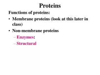
Outline • Biomédical Applications ofPlasma Gas Discharge/gamma irradiation Processes • Applications of Immobilizationof proteins onto polymer surfaces • Surface Properties Influencing Biological Responsesat Foreign Interfaces Possible modifications of an adsorbed protein layer.Radiation processed biomaterials Comparison of Biomaterials Coating Processes Examples: -The immobilized fungi for cellulase production - PVDF: Protein A ; Triglycine, Albumin, Fibrinogen, Immunoglobulin G - PLA: Lactoferrin
Biomedical Applications ofPlasma Gas Discharge/gamma irradiation Processes A. Plasma/Gamma irradiation Treatment (Etching) 1. Cleaning 2. Sterilization 3. Crosslink of surface layers B. Plasma/Gamma Irradiation Polymerization (Deposition) 1. Form Barrier Film a. Protective coating b. Insulating coating c. Reduce absorption from environment d. Reduce release rate of leachables e. Control drug delivery rate 2. Modify Protein and Cell Interactions a. Improve "biocompatibility" b. Promote selective protein adsorption c. Enhance cell adhesion d. Improve cell culture surfaces e. Provide non-fouling surfaces f. Reduce surface friction 3. Provide Reactive Sites a. For grafting or polymerizing polymers b. For immobilizing biomolecules
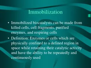
Applications of Immobilized Protein Biomolecules Organelles - any of a number of organized or specialized structures within a living cell.
Immobilization of proteins onto polymer surfaces – applications: • Developing medical implant materials; • Bioseparators; • Thin film fabrication; • Biochips and biosensors, • Drug delivery/drug release, • Food- and biochemical processing.
Possible modifications of the adsorbed protein layer with time after the initial layer is adsorbed.
E-enzyme NSA - N-succinimidyl acrylate
Immobilization of fungi and the continuous cell culture with the immobilized fungi for cellulase production Model scheme for the two kinds of immobilization methods for fungus (a) post immobilization method (b) in-source immobilization method
Protein molecules can be immobilized using chemical methods such as carbodiimide chemistry or sulfonyl chloride chemistry (sulfonyl chloride activates of hydroxylic materials) • Formation of mono- and multi-layer biomaterials on solid surfaces by using the Langmuir–Blodgett (LB) film and self-assembly layers techniques; • Grafting techniques; • Covalent immobilization of proteins; • Adsorption, cross-linking or covalent bonding, • Entrapment • Encapsulation
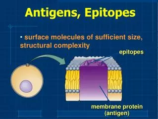
Biosensors • Essential tools in medicine, food quality control, environmental monitoring and research. • A biological molecule is immobilized in proximity to a transducer to detect an analyte, and ultimately it transduces the chemical signal produced by the interaction into a measurable response, most often an electronic signal
Diagram of a biosensor device Enzymes are by far the most commonly used biological components in biosensors. To be efficient they should be recovered.
Possibilities of proteins immobilization PEGylation or pegylation - process of both covalent and non-covalent attachment of polyethylene glycol Heparin or inulin
Substrates • PVDF – polyvinylidene fluoride • PLA – polylactic acid
PVDF surface functionalization by: • Radiofrequence plasma, microwave plasma treatment, electron beam, gamma irradiation, etc.; • Proteins Immobilization:
Q = 10 cm3/min, P = 50W t = 30 s d = 10 cm
Binding of protein is normally performed on a surface containing chemical groups reactive to the protein chemistry, like carboxylic acids or amines. • Depending on discharge gas, a large variety of chemical groups can be incorporated into the surface (e.g. hydroxyl, carbonyl, carboxylic, amino, or peroxyl groups). • Nitrogen, N2/H2 and ammonia plasma treatments give rise to N-containing functionalities, such as amine (−NH2), imine (−CH=NH), and nitrile (−C≡N) on polymer surfaces, as well as oxygen-containing groups, such as amides (−CONH2), due to post-discharge atmospheric oxidation – amphoteric, basic • Formation of carboxylic acids is favored mainly by the presence of the active species implemented by using CO2 as discharge gas - acidic. • To minimize the degradation effect on the biomaterial surface (since the formation of a degraded layer gives a weaker bond layer), the atomic species such as oxygen atoms in the plasma should be eliminated. • CO2 is less aggressive than oxygen and safer under working conditions. • Carboxyl groups are implemented also onto the gamma irradiated surfaces
Polyvinylidene fluoride films (PVDF) • Albumin • Triglycine (glycil-glycil-glycine)C6H11N2O4 • Protein A • Immunoglobulin G • Fibrinogen
AlbuminPhysical Adsorption ( Physisorption ) Chemical Adsorption ( Chemisorption ) Albumin bovine serum After plasma treatment
The albumin adsorption was done by dip-and-dry procedure by immersing the polymer films, at room temperature, in an aqueous solution of albumin of 2 wt%.
After coating the plasma pre-treated PVDF film surface with proteins, the hydrophilic character of the polymer increased. • As albumin-treated surfaces are resistant to platelet adhesion, an enhanced tolerance towards biological fluids and an increased haemocompatibility and biocompatibility characteristics of the polymer. • In the same time, the albumin increases the number of functional groups (−NH2 and −COOH) on the polymer surface, for binding other bioactive molecules, thus extending the polymer applicability.
Protein A and triglycine immobilization • A) Physisorption: protein solution made of protein A or triglycine was spread over the entire surface and stored at 4 ◦C overnight (for at least 15 h). • B) Chemisorption: The untreated and plasma-exposed surfaces were treated with EDC (1-ethyl-3-(-dimethylaminopropyl) carbodiimide) NHS (N-hydroxysuccinimide) at4 ◦C overnight for at least 15 h.
Covalent bonding of proteins in PVDF surface Acidic surface Amphoteric surface Basic Surface EDC - N-(3-Dimethylaminopropyl)-N′-ethylcarbodiimide hydrochloride; NHS- N-hydroxysuccinimide
Rough surfaces, whatever the nature of the treatment More smooth surfaces, especially with N2 or N2/H2 plasma activation and adsorption/grafting of proteins
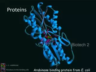
Immunofluorescence test results for virgin PVDF (a), N2/H2 plasma-treated PVDF with adsorbed TG (b), grafted with protein A (c) and grafted with TG (d). The proteins immobilized onto the PVDF surface exhibited the expected activity in coupling of the fluorescent antibody tracer as antiEscherichia coli, as assessed by an immunofluorescence test.
Plasma treatment of PVDF in a microwave plasma, followed by coating/grafting with different proteins, proved to be very useful for the appropriate modification of its surface properties, thus leading to a possible increase in the biocompatibility characteristics of the PVDF. • The proteins immobilized on the PVDF surface exhibited the activity in coupling of the fluorescent antibody tracer as anti Escherichia coli, as assessed by an immunofluorescence test. • Application: New piezoelectric biosensors for medical, aerospace, nuclearinstrumentation.
A piezoelectric sensor is a device that uses the piezoelectric effect, to measure changes in pressure, acceleration, temperature, strain, or force by converting them to an electrical charge. • They have been successfully used in various applications, such as in medical, aerospace, nuclear instrumentation
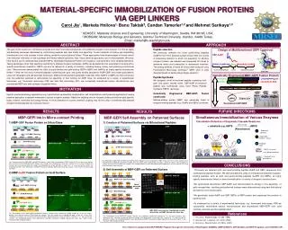
SAM Deposition of Immunoglobulin G via Protein A on PVDF radiation functionalized surface • IgG immobilization onto solid substrateis an important development in the imunosenzors. • Antibodies protect organism against infections by inactivation of viruses and patogen toxins; Layer-by-layer self-assembly (LbL) technique - it generates engineered nano-scale films, coatings, and particles used in multiple biomedical applications to create biosensors for antibody immobilization and detection. The multilayer system with protein-A has the potential to be further developed into an efficient immunoassay protein chip, when using piezoelectric PVDF. SAM- Self-assembled monolayers
Immunoglobulin G (IgG) is a type of antibody. It is a protein complex composed of four peptide chains—two identical heavy chains and two identical light chains arranged in a Y-shape typical of antibody monomers. Each IgG has two antigen binding sites.
Protein-A is specifically boundto the constant region of immunoglobulin, leaving the variableregions available for antigen bindingwhich results in oriented IgGimmobilization. When IgG is directly immobilized onto the PVDF surface, there are many possibilities for molecules to orient onto the surface instead, when using protein-A the number of possible orientation ways decreases and immobilization becomes more specific.
The content of IgG deposited onto the PVDF surface: • 3.2% ± 0.31 for directly immobilized IgG ; • 22.02% ± 0.13, for IgG immobilized via protein-A and the estimated content of protein-A was 11.96% ± 0.5. • 12.7% unknown component, occurring after the interaction with the polymeric substrate
Surface morphology IgG via protein A: • CO2 PrAads/IgG; (b) CO2 PrA leg/IgG; (c) N2 PrAads/IgG,; (d) N2 PrA leg/IgG; (e) N2/H2 PrAads/IgG; (f) N2/H2 PrA leg/IgG. • Surface roughness increases after plasma exposure higher for N2 și N2/H2plasma
Salmonella typhimuriumwas detected on surface by QCMtehnique.The antigen quantity absorbed in case of IgG immobilization via protein A is three times greater than that found in the case of direct immobilization. • Specific interaction antibody/antigen is improved by IgG immobilization via protein A • The self-assembled monolayer (SAM) obtained by immobilization of Immunoglobulin G onto PVDF surface in a highly oriented manner via Protein A has the potential to be used in the elaboration of new piezoelectric/immuno- biosensors (when using PVDF in β-phase, with piezoelectric properties).
Polyvinylidene fluoride (PVDF) film has been widely investigated as a sensor and transducer material due to its high piezo-, pyro- and ferroelectric properties. To activate these properties, PVDF films require a mechanical treatment, stretching or poling. • A force sensor based on PVDF fabrics with excellent flexibility and breathability, to be used as a specific human-related sensor. • Preliminary force sensors have been fabricated and demonstrated excellent sensitivity and response to external mechanical forces. This implies that promising applications for sensing garment pressure, blood pressure, heartbeat rate, respiration rate and accidental impact on the human body.
Fibrinogen • Protein adsorption is the first event taking place after implantation. The nature and composition of the first adsorbed protein layer is of major importance for the subsequent cellular response with blood or other tissues. • Fibrinogen is the major initiator of inflammatory response. Hence fibrinogen adsorption has to be controlled to prevent platelet adhesion and activation through adequate tailoring of surface properties like wettability and roughness. By tailoring the surface characteristics of the implant the adsorption of fibrinogen can be minimized. • It plays a vital role in thrombosis on surfaces. It is involved in the maintenance of heamostasis and serves as a ligand to a variety of vascular cells, including platelets, endothelial cells, and monocytes
FB contains two sets of three polypeptide chains (α, β, and γ). The simplest FB model comprises three spherical regions (one E domain and two D domains) connected by two narrow rods
CO2 Fb ads PVDF CO2 Fb leg Morphology and wettability • Roughness increases • Wetability is improved.
The presence of N, O and S on the PVDF surface demonstrate the fibrinogen grafting Contact angle decreased Roughness increases
FIBRINOGEN COATED PVDF - SCAFFOLD FOR CELL CULTURE Citotoxicityand cell viability evaluated on fibroblasts • Cellular viability is higher than 100% mainly for PVDF/N2/H2/FB/CC. • Cellular morphology is not modified. biomedical applications, especially for obtaining cell culture plates.
FB is covalently immobilized to the plasma-treated PVDF mainly through amide bonds. • The wettability of PVDF surface is improved by plasma treatment and FB immobilization, with a more pronounced hydrophilic character when using amphoteric and basic functionalization. • The obtained samples are not cytotoxic, have stimulated cellular division, and exhibit good surface properties. • Biomedical applications, especially for obtaining cell culture plates.
The protein immobilized PLLA may be widely used as a biocompatible material. • PLA with immobilized proteins - improvement of cellular interactions. Gelatin and collagen: To covalently immobilize gelatin or collagen type I on poly-L-lactic acid (PLLA) film surfaces poly(hydroxyethyl methacrylate) (PHEMA) or poly(methacrylic acid) (PMAA) was grafted via photooxidization and subsequent UVinduced polymerization. For films grafted with PHEMA, methyl sulfonyl chloride was used to activate the hydroxyl groups and for films grafted with PMAA and 1-ethyl-3-(3-dimethylaminopropyl) carbodiimide was used to activate the carboxyl groups. Gelatin and collagen were finally reacted with the activated hydroxyl or carboxyl groups to obtain covalently immobilized protein layers. Grafting of PHEMA, PMAA and protein on the surfaces was confirmed using ATR-IR and XPS. Surface wettability of the modified films was improved.
Substrate PLA Lactoferrin immobilization assisted by plasma discharge and gamma ray irradiation
LACTOFERIN is a multiple bioactive glycoprotein that is involved in several physiological functions, including: • regulation of iron absorption in the bowel; • immune response; • antioxidant, • anti carcinogenic and • anti-inflammatory properties; • protection against microbial infection, • Recommend it for utilization in obtaining new bioactive materials with different characteristics for human, animals and materials protection against microorganism attack
Chemical immobilization of the lactoferrin onto gamma irradiated PLA induces a roughness increase, which is associated with protein-clusters formation. Surface roughness with increasing the plasma treatment time or irradiation dose