Download
1 / 43
470 likes | 790 Views
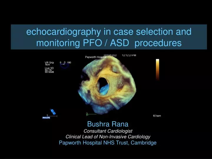
echocardiography in case selection and monitoring PFO / ASD procedures. Bushra Rana Consultant Cardiologist Clinical Lead of Non-Invasive Cardiology Papworth Hospital NHS Trust, Cambridge. overview. role of echocardiography in percutaneous atrial septum closure. ♥ pre-operative checklist

E N D
echocardiography in case selection and monitoring PFO / ASD procedures Bushra Rana Consultant Cardiologist Clinical Lead of Non-Invasive Cardiology Papworth Hospital NHS Trust, Cambridge
overview role of echocardiography in percutaneous atrial septum closure ♥ pre-operative checklist TOE protocol detailed assessment of defect ♥ TOE during the procedure some important considerations ♥ case examples highlighting important phenotypes
overview role of echocardiography in percutaneous atrial septum closure complexity of anatomy… crossing defect… device sizing … device positioning… complications…
pre-operative checklist tunnel length thickened secondary septum LA multiple openings STEP 1 confirm defect is secundum type STEP 2 surgery better option? anomalous pulmonary venous drainage mitral valve pathology STEP 3 detailed assessment of defect defect size number of defects surrounding rims atrial septal aneurysm STEP 4 interference during device placement? Eustachian valve and ridge Chiari network
transoesophageal echocardiography define anatomy assess suitability for device closure right atrium superior vena cava aortic root inferior vena cava coronary sinus tricuspid valve left atrium right upper pulmonary vein mitral valve SVC AO ASD CS IVC Rana et al, JACC CVI 2010; 3 (7): 749-60
TOE protocol 0 degrees transverse plane SVC AO high mid low FO CS IVC Rana et al, JACC CVI 2010; 3 (7): 749-60
TOE protocol 90 degrees longitudinal plane SVC high mid low AO FO CS IVC IVC Rana et al, JACC CVI 2010; 3 (7): 749-60
TOE protocol specialist viewsmid oesophagus AO 50 degrees SVC 120 degrees CS
detailed assessment of defect surrounding rims >5mm right atrium SVC (90-120) aortic root (50) IVC (90) CS (120) TV left atrium RUPV (0 &135) MV
detailed assessment of defect defect size…. PK McCarthy et al Im Paed Car 2003;15:1-24
detailed assessment of defect defect size…. PK McCarthy et al Im Paed Car 2003;15:1-24
detailed assessment of defect number of defects….how many holes are there? McCarthy et al Images Paediatr Cardiol 2003;15:1-24
detailed assessment of defect number of defects….how many holes are there? McCarthy et al Images Paediatr Cardiol 2003;15:1-24
detailed assessment of defect number of defects….how many holes are there? McCarthy et al Images Paediatr Cardiol 2003;15:1-24
detailed assessment of defect atrial septal aneurysm…mobility, margins need for over sizing to grip firm part of septum & ensure device stability
imaging during procedure wire position LAA/PV’s balloon inflation, defect sizing LA disc deployment RA disc deployment Position check working view 50 degrees
imaging during procedure wire position LAA/PV’s balloon inflation, defect sizing LA disc deployment RA disc deployment Position check Balloon inflation ♦with colour Doppler ♦ when flow stops balloon stops
imaging during procedure wire position LAA/PV’s balloon inflation, defect sizing LA disc deployment RA disc deployment Position check Balloon inflation ♦with colour Doppler ♦ when flow stops balloon stops ♦ at waist outer edge-outer edge
imaging during procedure wire position LAA/PV’s balloon inflation, defect sizing LA disc deployment RA disc deployment Position check Repeat protocol 0/50/90/120 ♦ device profile ♦ completely gripped septum ♦ abnormal flow
complex anatomy enface view from left atrium Rana et al Euro J Echocard 2010; 11: i19–i25
Ao RUPV MV
complex anatomy left atrium view Rana et al Euro J Echocard 2010; 11: i19–i25
complex anatomy left atrium view Rana et al Euro J Echocard 2010; 11: i19–i25
SVC FO ER CS IVC Rana et al Euro J Echocard 2010; 11: i19–i25
complex anatomy right atrium view Rana et al Euro J Echocard 2010; 11: i19–i25
complex anatomy RUPV MV Rana et al Euro J Echocard 2010; 11: i19–i25
complex anatomy LA view Rana et al Euro J Echocard 2010; 11: i19–i25
complex anatomy LA view Rana et al Euro J Echocard 2010; 11: i19–i25
complex anatomy LA view Rana et al Euro J Echocard 2010; 11: i19–i25
complex anatomy LA view Rana et al Euro J Echocard 2010; 11: i19–i25
defect position Rana et al Euro J Echocard 2010; 11: i19–i25
defect position Rana et al Euro J Echocard 2010; 11: i19–i25
summary complex anatomy… crossing defect… device sizing … device positioning… complications…
summary complex anatomy… crossing defect… device sizing … device positioning… complications…
role of echocardiography patient assessment for suitability of device closure diagnosis TTE ±bubble contrast detailed imaging intra-procedural guidance device sizing/position complications pre-procedural TOE intra-procedural TOE ICE GA Rana et al, JACC CVI 2010; 3 (7): 749-60
detailed assessment of defecttotal length of atrial septum Transverse 00 Longitudinal 900 mid oesophagus view mid oesophagus view