Download
1 / 57
580 likes | 883 Views
Reading the CXR. Frank Schembri Pulmonary / Critical Care. Types of Densities. Basic Principles of the CXR. Types of views PA Lateral AP Apical lordotic Decubitus (R & L). PA vs AP. Lateral CXR. Apical Lordotic Chest. Decubitus Positioning. Approaching the CXR.

E N D
Reading the CXR Frank Schembri Pulmonary / Critical Care
Basic Principles of the CXR • Types of views • PA • Lateral • AP • Apical lordotic • Decubitus (R & L)
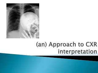
Approaching the CXR • Name, date, type of film • Type of film • Patient positioning / rotation • Inspiration • Penetration • White is underpenetrated • Black is overpenetrated
Approaching the CXR • The systematic approach 1. Tubes / Hardware 2. Bones 3. Soft tissues 4.Pleura and diaphragm 5. Trachea and mediastinum 6. Lung parenchyma
Lateral View Anterior View
Left Lung Right Lung
Lobes Right upper lobe:
Lobes (continued) Right middle lobe:
Lobes (continued) Right lower lobe:
Lobes (continued) Left lower lobe:
Lobes (continued) Left upper lobe with Lingula:
Lobes (continued) Lingula:
Lobes (continued) Left upper lobe - upper division:
Loss of volume Minor fissue Atelectasis mass Minor fissure Elevation of diaphragm
Minor fissure Major fissure
Pneumothorax • Collection of air in pleural cavity • Primary and secondary causes • Upright position air rises and separates the lung from the chest wall creating a line. Don’t be fooled by skin folds, clothing and bullae. • In the supine position air moves anteriorly. The lung will not be clearly separated from the chest wall.
Pneumothorax in the Supine Patient Enlarged hemithorax hyperlucent Mediastinal shift Deep sulcus sign Sharper cardiac border
Bat-winged appearance Enlarged heart