Download
1 / 72
790 likes | 1.2k Views
Chapter 8 Muscle Physiology. Outline. Structure Contractile mechanisms Mechanics Control Other muscle types Smooth, cardiac. Outline. Structure Muscle fiber (from myoblasts) Myofibrils Thick and thin filaments (actin and myosin) A,H,M,I,Z Sarcomere Titin –elasticity Cross bridges

E N D
Chapter 8 Muscle Physiology
Outline • Structure • Contractile mechanisms • Mechanics • Control • Other muscle types • Smooth, cardiac
Outline • Structure • Muscle fiber (from myoblasts) • Myofibrils • Thick and thin filaments (actin and myosin) • A,H,M,I,Z • Sarcomere • Titin –elasticity • Cross bridges • Myosin, Actin, tropomyosin, troponin
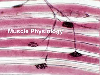
Muscle • Comprises largest group of tissues in body • Three types of muscle • Skeletal muscle • Make up muscular system • Cardiac muscle • Found only in the heart • Smooth muscle • Appears throughout the body systems as components of hollow organs and tubes • Classified in two different ways • Striated or unstriated • Voluntary or involuntary
Muscle • Controlled muscle contraction allows • Purposeful movement of the whole body or parts of the body • Manipulation of external objects • Propulsion of contents through various hollow internal organs • Emptying of contents of certain organs to external environment
Structure of Skeletal Muscle • Muscle consists a number of muscle fibers lying parallel to one another and held together by connective tissue • Single skeletal muscle cell is known as a muscle fiber • Multinucleated • Large, elongated, and cylindrically shaped • Fibers usually extend entire length of muscle
Muscle Tendon Muscle fiber (a single muscle cell) Connective tissue Fig. 8-2, p. 255
Structure of Skeletal Muscle • Myofibrils • Contractile elements of muscle fiber • Regular arrangement of thick and thin filaments • Thick filaments – myosin (protein) • Thin filaments – actin (protein) • Viewed microscopically myofibril displays alternating dark (the A bands) and light bands (the I bands) giving appearance of striations
Muscle fiber Dark A band Light I band Myofibril Fig. 8-2, p. 255
Structure of Skeletal Muscle • Sarcomere • Functional unit of skeletal muscle • Found between two Z lines (connects thin filaments of two adjoining sarcomeres) • Regions of sarcomere • A band • Made up of thick filaments along with portions of thin filaments that overlap on both ends of thick filaments • H zone • Lighter area within middle of A band where thin filaments do not reach • M line • Extends vertically down middle of A band within center of H zone • I band • Consists of remaining portion of thin filaments that do not project into A band
Z line A band I band Portion of myofibril M line H zone Sarcomere Thick filament A band I band Thin filament Cross bridges M line H zone Z line Myosin Actin Thick filament Thin filament Fig. 8-2, p. 255
I band A band I band Cross bridge Thick filament Thin filament Fig. 8-4, p. 256
Structure of Skeletal Muscle • Titin • Giant, highly elastic protein • Largest protein in body • Extends in both directions from M line along length of thick filament to Z lines at opposite ends of sarcomere • Two important roles: • Along with M-line proteins helps stabilize position of thick filaments in relation to thin filaments • Greatly augments muscle’s elasticity by acting like a spring
Myosin • Component of thick filament • Protein molecule consisting of two identical subunits shaped somewhat like a golf club • Tail ends are intertwined around each other • Globular heads project out at one end • Tails oriented toward center of filament and globular heads protrude outward at regular intervals • Heads form cross bridges between thick and thin filaments • Cross bridge has two important sites critical to contractile process • An actin-binding site • A myosin ATPase (ATP-splitting) site
Structure and Arrangement of Myosin Molecules Within Thick Filament
Actin • Primary structural component of thin filaments • Spherical in shape • Thin filament also has two other proteins • Tropomyosin and troponin • Each actin molecule has special binding site for attachment with myosin cross bridge • Binding results in contraction of muscle fiber
Actin and myosin are often called contractile proteins. Neither actually contracts. Actin and myosin are not unique to muscle cells, but are more abundant and more highly organized in muscle cells.
Tropomyosin and Troponin • Often called regulatory proteins • Tropomyosin • Thread-like molecules that lie end to end alongside groove of actin spiral • In this position, covers actin sites blocking interaction that leads to muscle contraction • Troponin • Made of three polypeptide units • One binds to tropomyosin • One binds to actin • One can bind with Ca2+
Tropomyosin and Troponin • Troponin • When not bound to Ca2+, troponin stabilizes tropomyosin in blocking position over actin’s cross-bridge binding sites • When Ca2+ binds to troponin, tropomyosin moves away from blocking position • With tropomyosin out of way, actin and myosin bind, interact at cross-bridges • Muscle contraction results
Cross-bridge interaction between actin and myosin brings about muscle contraction by means of the sliding filament mechanism.
Outline Contractile mechanisms • Sliding filament mechanism (Theory) • Ca dependence • Power stroke • T tubules • Ca release • Lateral sacs, foot proteins, ryanodine receptors, dihydropyradine receptors • Cross bridge cycling • Rigor mortis, relaxation, latent period
Sliding Filament Mechanism • Increase in Ca2+ starts filament sliding • Decrease in Ca2+ turns off sliding process • Thin filaments on each side of sarcomere slide inward over stationary thick filaments toward center of A band during contraction • As thin filaments slide inward, they pull Z lines closer together • Sarcomere shortens
Basic 4 steps Fig. 8-9, p. 260
Detailed steps Hydrolysis of ATP pivots head Release of ADP and Pi cocks head Fig. 8-13, p. 263
Power Stroke • Activated cross bridge bends toward center of thick filament, “rowing” in thin filament to which it is attached • Sarcoplasmic reticulum releases Ca2+ into sarcoplasm • Myosin heads bind to actin • Myosin heads swivel toward center of sarcomere (power stroke) • ATP binds to myosin head and detaches it from actin
Power Stroke • Hydrolysis of ATP transfers energy to myosin head and reorients it • Contraction continues if ATP is available and Ca2+ level in sarcoplasm is high
Sliding Filament Mechanism • All sarcomeres throughout muscle fiber’s length shorten simultaneously • Contraction is accomplished by thin filaments from opposite sides of each sarcomere sliding closer together between thick filaments
Relaxation • Depends on reuptake of Ca2+ into sarcoplasmic reticulum (SR) • Acetylcholinesterase breaks down ACh at neuromuscular junction • Muscle fiber action potential stops • When local action potential is no longer present, Ca2+ moves back into sarcoplasmic reticulum
T tubule Terminal button Surface membrane of muscle cell Acetylcholine- gated cation channel Lateral sacs of sarcoplasmic reticulum Acetylcholine Troponin Tropomyosin Actin Cross-bridge binding Myosin cross bridge Fig. 8-12, p. 262
Sarcoplasmic Reticulum • Modified endoplasmic reticulum • Consists of fine network of interconnected compartments that surround each myofibril • Not continuous but encircles myofibril throughout its length • Segments are wrapped around each A band and each I band • Ends of segments expand to form saclike regions – lateral sacs (terminal cisternae)
Transverse Tubules • T tubules • Run perpendicularly from surface of muscle cell membrane into central portions of the muscle fiber • Since membrane is continuous with surface membrane – action potential on surface membrane also spreads down into T-tubule • Spread of action potential down a T tubule triggers release of Ca2+ from sarcoplasmic reticulum into cytosol
Relationship Between T Tubule and Adjacent Lateral Sacs of Sarcoplasmic Reticulum
Outline • Mechanics • Tendons • Twitch • Motor unit • Motor unit recruitment • Fatigue • Asynchronous recruitment • Twitch, tetanus, summation • Muscle length, isometric, isotonic • Tension, origin, insertion
Skeletal Muscle Mechanics • Muscle consists of groups of muscle fibers bundled together and attached to bones • Connective tissue covering muscle divides muscle internally into bundles • Connective tissue extends beyond ends of muscle to form tendons • Tendons attach muscle to bone
Muscle Contractions • Contractions of whole muscle can be of varying strength • Twitch • Brief, weak contraction • Produced from single action potential • Too short and too weak to be useful • Normally does not take place in body • Two primary factors which can be adjusted to accomplish gradation of whole-muscle tension • Number of muscle fibers contracting within a muscle • Tension developed by each contracting fiber
Motor Unit Recruitment • Motor unit • One motor neuron and the muscle fibers it innervates • Number of muscle fibers varies among different motor units • Number of muscle fibers per motor unit and number of motor units per muscle vary widely • Muscles that produce precise, delicate movements contain fewer fibers per motor unit • Muscles performing powerful, coarsely controlled movement have larger number of fibers per motor unit
Motor Unit Recruitment • Asynchronous recruitment of motor units helps delay or prevent fatigue • Factors influencing extent to which tension can be developed • Frequency of stimulation • Length of fiber at onset of contraction • Extent of fatigue • Thickness of fiber
Twitch Summation and Tetanus • Twitch summation • Results from sustained elevation of cytosolic calcium • Tetanus • Occurs if muscle fiber is stimulated so rapidly that it does not have a chance to relax between stimuli • Contraction is usually three to four times stronger than a single twitch
Muscle Tension • Tension is produced internally within sarcomeres • Tension must be transmitted to bone by means of connective tissue and tendons before bone can be moved (series-elastic component) • Muscle typically attached to at least two different bones across a joint • Origin • End of muscle attached to more stationary part of skeleton • Insertion • End of muscle attached to skeletal part that moves
Types of Contraction • Two primary types • Isotonic • Muscle tension remains constant as muscle changes length • Isometric • Muscle is prevented from shortening • Tension develops at constant muscle length
Contraction-Relaxation Steps Requiring ATP • Splitting of ATP by myosin ATPase provides energy for power stroke of cross bridge • Binding of fresh molecule of ATP to myosin lets bridge detach from actin filament at end of power stroke so cycle can be repeated • Active transport of Ca2+ back into sarcoplasmic reticulum during relaxation depends on energy derived from breakdown of ATP