Download
1 / 51
530 likes | 735 Views
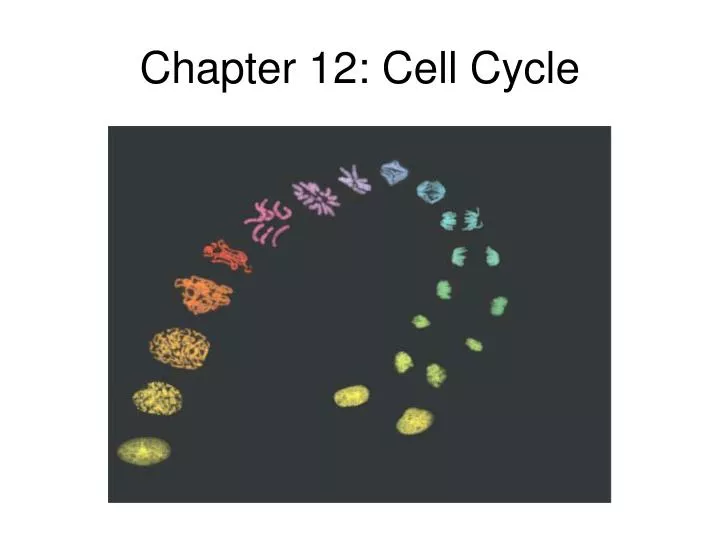
Chapter 12: Cell Cycle. Chromosome Sorting. The goal of cell division typically is to equally partition two more-or-less identical copies of genetic material between two daughter cells

E N D
Chromosome Sorting • The goal of cell division typically is to equally partition two more-or-less identical copies of genetic material between two daughter cells • Prokaryotes are comparatively simple, with only one chromosome, so have a relatively easy time sorting daughter chromosomes to daughter cells • Eukaryotes, with their longer DNA and multiple chromosomes, don’t have it nearly so easy • Much of the complex “dance” of Mitosis is a consequence of the need to make sure that each daughter cell ends up with the samenumber and type of chromosomes as the parent
Important roles of cellular division: Reproduction: Forms duplicate offspring (amoeba). Growth and Development: Allows single cell to form into multicelluar organism Tissue Renewal: Cells are damaged and die all the time. These cells need to be replaced. Cell Division
Chromosomal Differences Prokaryote Chromosome Eukaryote Chromosome Here “chromosome” and “DNA” are not 100% synonymous
Chromosomes Tightly packaged DNA Found only during cell division DNA is not being used for macromolecule synthesis Chromatin Unwound DNA Found throughout Interphase DNA is being used for macromolecule synthesis Chromosomes vs. Chromatin
Eukaryotic Chromosomes Though chromosomes are “all about” DNA, in fact much this structure consists of protein Formed via replication, not by formed chromatids coming together
A chromatid is a chromatid as long as it is held in association with a sister chromatid at the centromere When two sister chromatids separate (after metaphase) they go from being a single chromosome to being two different chromosomes How Long is a Chromatid?? Sister Chromatids Chromatid Chromosome
Eukaryotic Chromosome Genome = DNA Chromosomes = DNA + PROTEIN (visible under light microscope) Chromatin = DNA + PROTEIN (unwound) DNA
Important Vocabulary • Centromere • Centrosome • Centriole • Kinetochore • Kinetochore microtubules • Mitotic spindle • Nonkinetochore microtubules • Spindle apparatus • Spindle fibers
Important Vocabulary 2 Centrioles = Centrosome • Centromere • Centrosome • Centriole • Kinetochore • Kinetochore microtubules • Mitotic spindle • Nonkinetochore microtubules • Spindle apparatus • Spindle fibers Nonmembranous organelles that organize microtubules throughout the cell cycle
Important Vocabulary • Centromere • Centrosome • Centriole • Kinetochore • Kinetochore microtubules • Mitotic spindle • Nonkinetochore microtubules • Spindle apparatus • Spindle fibers Comprised of microtubules. Only in animal cells! Not necessary for spindle formation.
Important Vocabulary • Centromere • Centrosome • Centriole • Kinetochore • Kinetochore microtubules • Mitotic spindle • Nonkinetochore microtubules • Spindle apparatus • Spindle fibers Attachment point on Chromatids for spindle fiber
Important Vocabulary • Centromere • Centrosome • Centriole • Kinetochore • Kinetochore microtubules • Mitotic spindle • Nonkinetochore microtubules • Spindle apparatus • Spindle fibers The portion of the mitotic spindle that is connected to the chromosome during mitosis
Important Vocabulary • Centromere • Centrosome • Centriole • Kinetochore • Kinetochore microtubules • Mitotic spindle • Nonkinetochore microtubules • Spindle apparatus • Spindle fibers The microtubules that are responsible for separating as well as pushing centrosomes towards the opposite ends of the cells.
Important Vocabulary • Centromere • Centrosome • Centriole • Kinetochore • Kinetochore microtubules • Mitotic spindle • Nonkinetochore microtubules • Spindle apparatus • Spindle fibers Microtubules of the mitotic spindle that are not connected to chromosomes but are responsible for pushing centrosomes apart.
Important Vocabulary • Centromere • Centrosome • Centriole • Kinetochore • Kinetochore microtubules • Mitotic spindle • Nonkinetochore microtubules • Spindle apparatus • Spindle fibers The mitotic spindle as visible through a light microscope.
Important Vocabulary • Centromere • Centrosome • Centriole • Kinetochore • Kinetochore microtubules • Mitotic spindle • Nonkinetochore microtubules • Spindle apparatus • Spindle fibers Bundles of microtubules that comprise the spindle apparatus. This bundling is what allows us to visualize the fibers through a light microscope
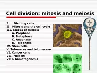
Interphase (not mitosis) Mitosis Prophase Prometaphase Metaphase Anaphase Telophase Cytokinesis Phases of the Cell Cycle (Eukaryotes)
Interphase Mitosis is the shortest part of the cell cycle. Interphase accounts for about 90% of the cell cycle. Cells: Grow Copy chromosomes Prepare for cell division Phases of the Cell Cycle (Eukaryotes)
Interphase is divided into subphases: G1 Phase = Gap 1 S Phase = synthesis G2 Phase = Gap 2 During all three subphases, the cell grows by producing proteins and organelles. Phases of the Cell Cycle (Eukaryotes)
Figure 12.5 The stages of mitotic cell division: interphase Late interphase • Nucleus is well defined and bounded by the nuclear envelope • Duplicated chromosomes are loosely packed chromatin fibers • Microtubules extend from the duplicated centrosomes
Figure 12.5 The stages of mitotic cell division: prophase Prophase: 1st Phase of Mitosis • Nucleoli disappear • Chromatin fibers become tightly coiled in the nucleus condensing into discrete chromosomes • Centrosomes move away from each other as mitotic spindle begins to form in the cytoplasm
Figure 12.5 The stages of mitotic cell division: prometaphase Prometaphase • Nuclear envelope fragments • Chromosomes condense further and form kinetochores (structures at the centromere region that microtubules bind) • Microtubules extend from each pole and invade the nuclear area and either attach to kinetochores or microtubules from the opposite side
Figure 12.5 The stages of mitotic cell division : metaphase Metaphase • Centrisome are at opposite poles • Chromosomes line up at the center of the cell equidistant from each pole (metaphase plate) • Microtubules are attached to the kinetochores of each sister chromatid facing its pole
Figure 12.5 The stages of mitotic cell division : anaphase Anaphase • Paired centromeres of each chromosome separate, dividing the sister chromatids • Centrosome poles move farther apart • Microtubules begin to shorten, pulling their attached chromosome towards opposite poles
Figure 12.5 The stages of mitotic cell division : telophase and cytokinesis Telophase • Daughter nuclei form at the two poles • Nuclear envelopes begin to form from the fragments of the parent cell • Chromatin fibers loosen Cytokinesis • A cleavage furrow forms between daughter cells and the cell is pinched in two, equally dividing the cytoplasm
Figure 12.5x Mitosis Prometaphase
Aster Centrosome MetaphasePlate Sisterchromatids Kinetochores Overlappingnonkinetochoremicrotubules Kinetochores microtubules 0.5 µm Microtubules Chromosomes Figure 12.7 Centrosome 1 µm The Mitotic Spindle: A Closer Look • The mitotic spindle • Is an apparatus of microtubules that controls chromosome movement during mitosis • The spindle arises from the centrosomes • And includes spindle microtubules and asters • Some spindle microtubules • Attach to the kinetochores of chromosomes and move the chromosomes to the metaphase plate
Separating Chromosomes • During anaphase, sister chromatids are separated • Proteins that hold the chromatids together are inactivated • Kinetochores have motor proteins that move the chromosomes along microtubules towards the spindle poles • Nonkinetochore microtubules elongate the cell
Figure 12.7 Testing a hypothesis for chromosome migration during anaphase Model: Chromosomes travel along microtubules towards the poles Microtubules shorten by depolymerizing at their kinetochore ends Experiment: Microtubules of dividing cells are labeled with a fluorescent dye A laser bleaches the dye in a region midway between one spindle pole and the chromosome As chromosomes move towards the poles, microtubules on the kinetochore side of the mark shortened, while those on the centrosome side remained the same length
Cytokinesis : division of cytoplasm • Animal cells : cleavage • Cleavage furrow • Division begin as a shallow groove in the cell surface near the metaphase plate • Contractile ring (on the cytoplasmic side of the furrow) • Composes of actin microfilaments and the protein myosin • Cleavage furrow deepens until the parent cell is pinched in two • Plant cells : cell plate formation • Vesicles from the Golgi collect at the middle of the cell producing a cell plate • Cell wall material carried in the vesicles is deposited on the plate as it grows, until it fuses with the membrane along the perimeter of the cell
Figure 12.9 Mitosis in a plant cell Chromatin condensing Spindle forms Nuclear envelope fragments Cell plate forms Microtubules capture kinetochores Chromosomes line up at metaphase plate Chromatids separate and move towards poles
Evolution of Mitosis: prokaryotic reproduction • Cell division by binary fission • Bacterial chromosome is a single circle of DNA • Replication of DNA begins at a specific origin of replication • Duplicated chromosomes actively move apart without the help of mitotic spindle
Figure 12.11 A hypothesis for the evolution of mitosis Chromosomes move to opposite ends of the cell by unknown mechanisms Microtubules pass through the nucleus in cytoplasmic tunnels Nuclear envelope remains intact Chromosomes attach to envelope Protists: Plankton Mitotic spindle form in the nucleus Nuclear envelope remains intact Microtubules separate the chromosomes Algae: Phytoplankton Mitotic spindle form outside of the nucleus Nuclear envelope breaks down Microtubules separate the chromosomes
Cell Cycle Regulation • Timing and rate of cell division is critical for normal growth, development, and maintenance • Skin cells divide frequently • Liver cells divide only when needed (repair) • Muscle cells and nerve cells do not divide • Molecular mechanisms regulate the cell cycle • Improper cell cycle regulation can result in human disease : cancer
EXPERIMENTS In each experiment, cultured mammalian cells at two different phases of the cell cycle were induced to fuse. Experiment 2 Experiment 1 S G1 M G1 RESULTS M S S M When a cell in the S phase was fused with a cell in G1, the G1 cellimmediately entered the S phase—DNA was synthesized. When a cell in the M phase was fused with a cell in G1, the G1 cell immediately began mitosis— a spindle formed and chromatin condensed, even though the chromosome had not been duplicated. CONCLUSION The results of fusing cells at two different phases of the cell cycle suggest that molecules present in the cytoplasm of cells in the S or M phase control the progression of phases. Figure 12.13 A, B Evidence for Cytoplasmic Signals • Molecules present in the cytoplasm • Regulate progress through the cell cycle
Figure 12.13 Mechanical analogy for the cell cycle control system The cell cycle is regulated at certain checkpoints by both internal and external controls
Checkpoints • Critical regulatory points where activating and inhibiting signals can control progression through the cell cycle • Stop signals predominant at checkpoints until overridden by an activating signal • Signals come from cell surveillance mechanisms • Informing the cell when all processes in the current phase have been completed correctly or not • Signals come from outside the cell • Checkpoints • G1 phase : restriction point • cells that do not pass this point go into a nondividing G0 phase • G2 phase • M phase
Cell Cycle Control Molecules • The fluctuation in the amount and activity of regulatory molecules in the cytoplasm pace the sequential events of the cell cycle • Kinases that drive the cell cycle - Cdks (cyclin dependent kinases) • Present at a constant concentration in an inactive form • Activated by attachment to a cyclin • Activity of Cdks rises and falls with changes in the concentration of its cyclin partner
Figure 12.14 Molecular control of the cell cycle at the G2 checkpoint • MPF = M phase (maturation) Promoting Factor • triggers the passage past the G2 checkpoint into M phase by phosphorylating proteins involved in nuclear envelope breakdown • Initiates process leading to the destruction of its cyclin, switching itself off and driving the cell past the M phase checkpoint MPF causes the breakdown of cyclin MPF promotes mitosis Cdk + cyclin combine to form MPF
Signals that Regulate the Cell Cycle • Internal signals • M phase checkpoint: anaphase does not begin until all chromosomes are attached to spindle on the metaphase plate • Kinetochores not attached to spindle send a signal to delay anaphase • External signals • Growth factors (eg: PDGF) • Density-dependent inhibition of cell division
Figure 12.15 The effect of a growth factor on cell division Platelet-derived growth factor (PDGF) stimulates the division of human fibroblast cells
Figure 12.16 Density-dependent inhibition of cell division Density-dependent inhibition: A cell population reaches a certain density, growth factors and nutrients available to each cell becomes insufficient to allow continued cell growth Anchorage dependence: Cells must be attached to a substratum to divide – signals are transmitted to cell cycle control via plasma membrane proteins and elements of the cytoskeleton linked to them
Cancer • Cancer cells do not respond to normal cell cycle controls and divide excessively • Make a required growth factor themselves • Abnormal cell cycle control system • Abnormality in signaling pathway that conveys the growth factor signal • Cancer cells divide indefinitely if supplied with nutrients: in vitro cell lines (HeLa cells) • Transformation – the process that converts a normal cell to a cancer cell • Tumor – a mass of abnormal cells that have evaded the immune system • Benign: cell mass remains at original site • Malignant: tumor invades organs and impairs function • Metastasis – spread of cancer cells to locations distant from the original site • cancer cells can lose attachments to other cells and spread into nearby tissues or enter the blood stream.
Figure 12.17 The growth and metastasis of a malignant breast tumor