Download
1 / 143
1.43k likes | 1.44k Views
Nervous System. What allows you to perceive the world around you, to recognize the incoming stimuli and to react to the environment Sensory systems Motor systems Memory/learning. Nervous System. Central Nervous System (CNS) Brain and Spinal Cord Peripheral Nervous System (PNS)

E N D
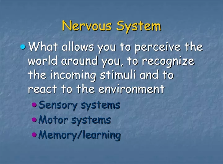
Nervous System • What allows you to perceive the world around you, to recognize the incoming stimuli and to react to the environment • Sensory systems • Motor systems • Memory/learning
Nervous System • Central Nervous System (CNS) • Brain and Spinal Cord • Peripheral Nervous System (PNS) • All nervous tissue outside the CNS • Somatic Nervous System (SNS)---voluntary • Sensory and motor neurons to skeletal muscle • Autonomic Nervous System (ANS)---Involuntary • Sensory and motor neurons to viscera • Sympathetic Division (increase heart rate) • Parasympathetic Division (decrease heart rate)
Nervous System Neurons (=nerve cells) general characteristics: - can be very long---6 feet or more - conduct nervous impulses---communication - long lived---entire life span - high metabolic rate; high O2 and glucose needs---brain accounts for less than 10% of body mass, yet gets over 25% of body’s energy
Types of Neurons (functional classification) • Motor Neurons--- • carry info from brain to body • Sensory Neurons--- • carry info from body to brain • Interneurons--- • carry info within the Central Nervous System (CNS). These account for 90% of the neurons in the body • Neuroglia---support cells • Oligodendrocytes---CNS • Schwann cells---PNS
Types of Neurons (structural classification) • Multipolar---many processes located in CNS • Bipolar---2 processes located in sensory organs (eye, ear & nose) • Unipolar--1 process these are sensory neurons. The axon and dendrite fuse into one process. • Interneurons---Named based on looks or after the person who first described them (fig. 12.5) • Purkinje cells, Pyramidal cells
Anatomy of a `typical’ motor neuron nucleus cell body = soma = perikaryon
Anatomy of a `typical’ motor neuron nucleus Nissl bodies = rough endoplamic reticulum cell body = soma = perikaryon
Anatomy of a `typical’ motor neuron nucleus Nissl bodies cell body = soma = perikaryon Golgi apparatus
Anatomy of a `typical’ motor neuron other organelles: mitochondria lysosomes neurofilaments nucleus Nissl bodies cell body = soma = perikaryon Golgi apparatus
Anatomy of a `typical’ motor neuron neural processes: axon = nerve fiber dendrites
Anatomy of a `typical’ motor neuron Flow of Information: axon dendrites
Anatomy of a `typical’ motor neuron axon hillock (trigger zone) axon dendrites
Anatomy of a `typical’ motor neuron terminal branches= telodendria axon hillock axon dendrites
Anatomy of a `typical’ motor neuron terminal branches axon hillock axon axonal terminals = synaptic knobs = synaptic boutons dendrites
Anatomy of a `typical’ motor neuron terminal branches axon hillock axon axonal terminals = synaptic knobs = synaptic boutons dendrites
Anatomy of a `typical’ motor neuron axonal terminals = synaptic knobs = synaptic boutons
Anatomy of a `typical’ motor neuron synaptic vesicles with neurotransmitters
Myelin Sheath • Some Axons are myelinated • Myelin Sheath (schwann cell in PNS and oligodendrocyte in CNS) axon • Multiple layers wrapped around = myelin • Cell body and cytoplasm (outer most layer) = Neurolemma
Anatomy of a `typical’ motor neuron myelination:
Anatomy of a `typical’ motor neuron myelination: Special Glial cells -- make up the myelin sheath
Anatomy of a `typical’ motor neuron nodes of Ranvier
Resting Membrane Potential • Voltage across the membrane • Electric potential energy • Due to separation of ions • All cells have a resting membrane potential
- + + + + - - + + - - - Resting Membrane Potential - + + + + - - + + - - -
Resting Membrane Potential + - + - - + - - - + + +
Amplifier Electrode 0 mV Oscilloscope Resting Membrane Potential
Amplifier Electrode -70 mV Oscilloscope Resting Membrane Potential
Resting Membrane Key Players • Sodium-Potassium (Na+/K+) Pump • Moves Sodium and Potassium against their concentration gradients • Moves 3 Na+ out of the cell and 2 K+ into the cell
Resting Membrane Potential Key Players: Na+/ K+ Pump: 3 Na+ out Cell exterior cell membrane ATP Cell interior 2 K+ in
Resting Membrane Potential Key Players: Leaky Channels Na+ K+ Cell exterior cell membrane K+ Cell interior Na+ • Many K+ “leak” channels • Few Na+ “leak” channels
ATP ATP _ + _ + _ + ATP _ + _ _ + _ _ + + + K+ 2 K+ K+ K+ 2 K+ Pumps 3 Na+ Leak Channels K+ 2 K+ K+ K+ 3 Na+ Na+ Together creates -70 mV Resting Membrane Potential 3 Na+ 2 K+ Na+ Leak Channels Na+
Resting Membrane Potential • Voltage across the membrane • Electric potential energy • Due to separation of ions • All cells have a resting membrane potential
Electrical Signaling Through Changes in Membrane Potential • Describing Changes in Membrane Potential • Graded Potentials • Action Potentials • Propagation of Action Potentials
0 mV -70 mV -90 mV 0 mV -70 mV -90 mV Two Types of Change: • Depolarizing • reduce membrane potential • make inside less negative • Na+ move into cell • Hyperpolarizing • increase membrane potential • make inside more negative • K+ move out of cell
Signals produced by a change in membrane potential • Graded potentials • Short lived • Local Signaling • Either depolarizing or hyperpolarizing
Graded Potentials • typical of dendrites and cell body (NOT axons) • are the first step in neuronal communication • are essential in initiating action potentials