Download
1 / 127
1.38k likes | 1.79k Views
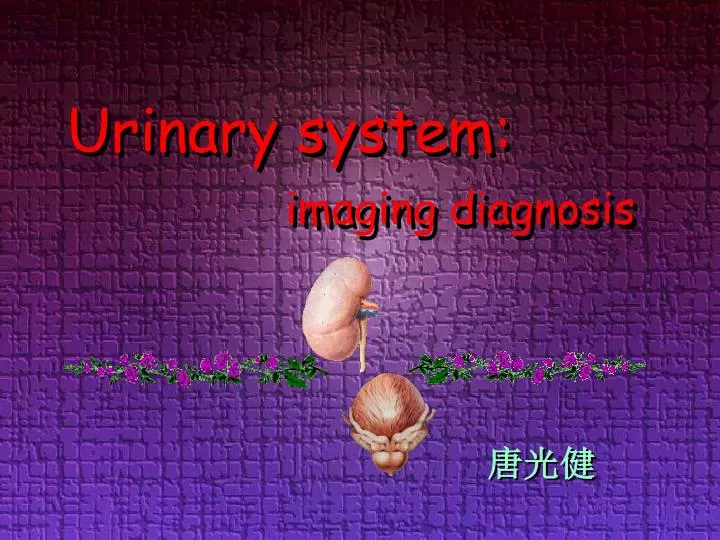
Urinary system: imaging diagnosis. 唐光健. Questions to think of. Which diagnostic imaging modality is the most suitable for the urinary system and why? What urinary diseases are suitable for imaging examination?

E N D
Questions to think of • Which diagnostic imaging modality is the most suitable for the urinary system and why? • What urinary diseases are suitable for imaging examination? • What imaging method should be chosen with different sort of disease considered clinically? What information we expect to get from the imaging?
Urinary System Characteristics: poor natural contrast • Luminal Structure • calyces, pelvic, ureter, bladder -UROGRAPHY • Parenchyma • Kidney-cortex, medulla -TOMOGRAPHIC IMAGING
Imaging modalities: • plane film(KUB)-limited effective • Urography • Intravenous pyelography IVP • Retrograde pyelography • Cystography • Urethrography -show morphous of the lumen indirectly -overall view of the urinary tract -affected by the renal function & lumen construction • Ultrasonography USG-real time, convenient, low cost, available -good characteristic of stone, fluid • CT Computed Tomography -high resolution of high density & low density -contrast enhancement • MRI Magnetic Resonant Imaging -good contrast of soft tissues
Normal Manifestations • KUB: contour of kidney -edge, density, size, position…
Normal Manifestations • IVP –compress the ureter, picture 15’-30’ after contrast administration renalpapilla Vtx body formix major calyx-neck fundament minor calyx renal pelvic ureter bladder
Normal Manifestations • IVP –compress the ureter, picture 15’-30’ after contrast administration renalpapilla Vtx body formix major calyx-neck fundament minor calyx renal pelvic ureter
Normal Manifestations • IVP-normal variant branching ampullary
Normal Manifestations • retrograde pyelogram back flow-collecting duct BF, renal sinus BF, perivein BF, lymph duct BF
Normal Manifestations • B-mode sonography- renal sinus, renal parenchyma, renal profile…
Normal Manifestations • CT– without/with contrast ~cortical phase, parenchymal phase, excrete phase ~profile, density, enhancement… Parenchymal phase cortical phase plane scan
Normal Manifestations • CT– without/with contrast ~cortical phase, parenchymal phase, excrete phase ~profile, density, enhancement… Parenchymal phase excretive phase cortical phase
Normal Manifestations • Angiography– ~no routine method, with interventional therapy ~arterial ph. parenchymal ph. Veinal ph.
Normal Manifestations • MRI- T1WI higher SI with cortex T2WI homogeneous high SI (signal intensity) T2WI T1WI
Renal pathologies • pathologies of Parenchymal • Solid -benign: angiomyolipoma -malignant: carcinoma • Cystic inside kidney: renal cyst outside kidney: sub capsule hematoma • pathologies of Urinary tract • of the wall –transitional cell carcinoma • of the lumen –calculus • Inflammatory change-tuberculosis • Hydronephrosis –differentiate diagnosis
Renal pathologies • pathologies of Parenchymal • Solid -benign: angiomyolipoma -malignant: carninoma • Cystic inside kidney: renal cyst outside kidney: sub capsule hematoma • pathologies of Urinary tract • of the wall –transitional cell carcinoma • of the lumen –calculus • Inflammatory change-tuberculosis • Hydronephrosis –differentiate diagnosis
Renal solid malignant • renal carcinoma - neoplasm, vascularity, necrosis
Renal solidmalignant • pathologic features correlate to imaging • neoplasm-mass effect • neoplastic vascularity • necrosis & bleeding in side the mass
Renal solidmalignant • renal carcinoma - neoplasm, vascularity, common with necrosis • plain film: profile change-helpless with diag. • urography: elongated & deformed pelvic- poor specification • BUS: substantial, hypo/inhomogeneous echoic • angiography: vessel shift, tumor flashing, neo-vessel. Staging • CT high specific & sensitive. Staging • MRI Seldom used
Renal solidmalignant • renal carcinoma • plain film: profile change-helpless with diag.
Renal solidmalignant • renal carcinoma • urography: elongated & deformated pelvic-poor specification
Renal solidmalignant • renal carcinoma • BUS: substantial, hypo/inhomog eneous echoic
Renal solidmalignant • renal carcinoma • CT high specific & sensitive. Staging plain scan delay scan With enhancemnet
Renal solidmalignant • renal carcinoma • CT high specific & sensitive. Staging delay scan plain scan with enhancement
Renal solidmalignant parenchymal phase • renal carcinoma • angiography: vessel shift, tumor flashing, neo-vessel. Staging post ia adrenalin arterial phase
Renal solidmalignant • renal carcinoma • CT high specific & sensitive. Staging renal vein, IVC invaded by RCC
Renal solidmalignant • renal carcinoma • CT high specific & sensitive. Staging renal vein, IVC invaded by RCC
Renal solidmalignant • renal carcinoma • angiography: vessel shift, tumor flashing, neo-vessel. Staging Arterial phase Parenchymal phase
Renal s solidmalignant • renal carcinoma • angiography: vessel shift, tumor flashing, neo-vessel. Staging post embolizing of RA IVC angiography
Renal solidmalignant • renal carcinoma • MRI seldom use
Renal solidbenign • Renal hamartoma (angiomyolipoma)- ratio of tree elements various, bleeding often
Renal solidmalignant Angiomyolipoma • pathologic features correlate to imaging • neoplasm-mass effect • adipose tissue, smooth muscle, vessels • hypo-developed neoplastic vessels-bleeding
Renal solidbenign • renal angiomyolipoma • plain film: profile change-helpless with diag. • urography: elongated & deformed pelvic-poor specification • sonography: hyperechoic substantial mass mostly • CT: high specific with fat density, high sensitive, bleeding might be seen • MRI inhomogeneous signal, signal of fat & old bleeding
Renal solidbenign • renalangiomyolipoma • sonography: hyperechoic substantial mass mostly
Renal solidbenign CT plain scan • renalangiomyolipoma • CT high specific with fat density, high sensitive, bleeding might be seen with enhancement
Renal solidbenign • renalangiomyolipoma • CT high specific with fat density, high sensitive, bleeding might be seen with enhancement CT plain scan
Renal solidbenign • renalangiomyolipoma • CT high specific with fat density, high sensitive, bleeding might be seen with enhancement Coronal MPR
Renal pathologies • pathologies of Parenchymal • Solid -benign: angiomyolipoma -malignant: carninoma • Cystic inside kidney: renal cyst outside kidney: sub capsule hematoma • pathologies of Urinary tract • of the wall –transitional cell carcinoma • of the lumen –calculus • Inflammatory change-tuberculosis • Hydronephrosis –differentiate diagnosis
Renal pathologies • pathologies of Parenchymal • Solid -benign: angiomyolipoma -malignant: carninoma • Cystic inside kidney: renal cyst outside kidney: sub capsule hematoma • pathologies of Urinary tract • of the wall –transitional cell carcinoma • of the lumen –calculus • Inflammatory change-tuberculosis • Hydronephrosis –differentiate diagnosis
cystic, inside kidney-renal cyst cyst • pathologic features correlate to imaging • with thin, even wall, • flat epithelium • clear fluid inside & no blood flow
cystic, inside kidney-renal cyst • plain film: profile changing • urography: pelvix & calix deforming, no specific • BUS: anechoic lesion, high specific • CT: clear appearance, high sen. & spe. for calcification of the wall or septation • MRI:cystic, signal of fluids, different diagnosis needed if signal be non-specific
cystic, inside kidney-renal cyst • Urography deforming of pelvic & calculi, low specific
cystic, inside kidney-renal cyst C • BUS, high specific anechoic lesion, echo enhancement post.
cystic, inside kidney-renal cyst C • CT: clear appearance, high sen. & spe. for calcification of the wall or septation Waterlike, no enhancement, “no wall”
cystic, inside kidney-renal cyst C • CT: clear appearance, high sen. & spe. for calcification of the wall or septation
cystic, inside kidney-renal cyst C • CT: clear appearance, high sen. & spe. for calcification, differentiation with high density cyst Enhancement scan Plain scan
cystic, inside kidney-renal cyst C • MRI, atypical signal-differentiate cystic, sig. of fluid
cystic, inside kidney-renal cyst Multicystic kidney
cystic, inside kidney-multycystic kidney • plain film: enlargement & shift of renal shadow • urography: elongation/deformation of the pelvic • BUS: renal enlargement of both sides with multi-cyst ~ diagnosed • CT: manifest clear, with multi-cystic liver often
cystic, inside kidney-multycystic kidney • plain film: enlargement & shift of renal shadow • urography: elongation/deformation of the pelvic • BUS: renal enlargement of both sides with multi-cyst ~ diagnosed • CT: manifest clear, with multi-cystic liver often