Download
1 / 44
440 likes | 451 Views
Learn about the significance of cell division in living organisms, including its role in growth, repair, and reproduction. Discover the process of mitosis and the importance of chromosomes in cell division.

E N D
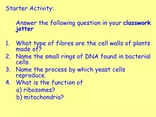
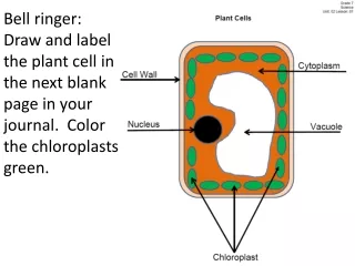
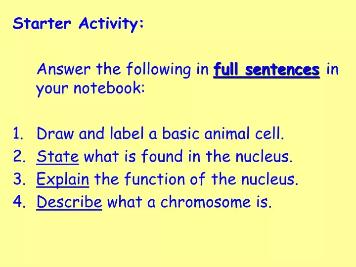
Starter Activity: Answer the following in full sentencesin your notebook: Draw and label a basic animal cell. State what is found in the nucleus. Explain the function of the nucleus. Describe what a chromosome is.
Key Area 3 Making new cells
Making New Cells Learning Intention: To understand how living organisms grow. Success Criteria: State where genetic material is found in living organisms. Understand the need for cell division. Explain the process of mitosis. Give uses of cell division.
Importance of Cell Division • In animals, this cell is formed when an egg cell is fertilised by a sperm cell. • The fertilised egg then divides into many cells to form an embryo. • The embryo continues to grow into a new animal.
Making New Cells • New cells are made to allow living organisms to grow. • New cells are also made to replace dead cells, repair tissues and heal wounds. • All new cells are made from existing cells by the process of cell division. Cell division definition - Twig clip
Importance of Cell Division pollen grain • Every living organism produced by sexual reproduction starts life as one cell. • In plants, this cell is formed when an egg cell is fertilised by a pollen grain. • The fertilised egg cell then divides into many cells to form a seed. • The seed can then grow into a new plant. pollen tube pollen nucleus egg cell
Growth • All living organisms grow during their lifetime. • All of this growth happens due to cells dividing to form new cells.
Repair to tissues • When we get injured, new cells are made to repair and replace the damaged tissues. • New skin cells form to heal a cut or graze, and new bone cells grow to repair a broken bone.
Cell Division • The process of cell division is called mitosis. • Mitosis is required for growth and repair.
Importance of Cell Division Your task.. Think: On a show me board list ideas why cell division is important. Pair: Decide with your partner two key reasons why cell division is important. Share: Show your ideas on one show me board In your notebook: List the key reasons why cell division is important.
Making New Cells Learning Intention: To understand how living organisms grow. Success Criteria: State where genetic material is found in living organisms. Understand the need for cell division. Explain the process of mitosis. Give uses of cell division.
Starter Activity: On a show me board: Why is cell division important?
Cells and chromosomes • Inside the nucleus of a cell there are chromosomes which carry the genetic information of the organism. • Each chromosome is made up or two chromatids. • Human cells have 46 chromosomes in their nucleus.
Matching sets of chromosomes • Normal cells have 2 sets of matching chromosomes. • These are known as diploid cells. • In humans this is 2 sets of 23 chromosomes. • This diagram shows the chromosomes of a human female laid out in order of size.
Cell division • Cells are able to make new cells by cell division. • The parent cell splits to form two cells. • Each new cell is identical to the original parent cell. Embryonic cell division
Chromosomes and cell division Spindle fibres pull the chromatids apart. • The chromosomes all make copies of themselves in a process called DNA replication. • The cell then divides into two diploid cells. This is called mitosis. • Each new cell ends up with the same number of chromosomes as the parent cell. • This means each new cell has all the genetic information. Twig Video Clip - Cell Division: Mitosis
Mitosis in detail Spindle fibres Chromosomes line up on the equator of the cell Equator (middle) Each chromosome duplicates to make a double chromosome Chromosomes split into two and spindle fibres pull the chromatids apart Cytoplasm divides to make two cells
Looking at dividing cells – root tip slides Equipment : microscope, prepared slide of onion root tip Method : • Set up the microscope at low power. • Look at the area just behind the tip of the root at low power. • Look for cells where chromosomes can be seen. • Increase to medium and then high power. • Look for cells where the chromosomes are about to separate, and where they have already separated. • Make enlarged drawings of three different cells.
Mitosis in action • You will use pipe cleaners and card to make a model of a cell. • You will then take the cell through the process of mitosis. • Your teacher will lead you through this activity.
Making New Cells Learning Intention: To understand how living organisms grow. Success Criteria: State where genetic material is found in living organisms. Understand the need for cell division. Explain the process of mitosis. Give uses of cell division.
Starter Activity: In pairs: Describe the process of mitosis
Starter 2
Modelling Mitosis We must… Plan and prepare mitosis video clips. We should… Swap and evaluate each other’s clips. We could… Use feedback to improve the clips.
Modelling Mitosis The mitosis video clip must: • be at least one minute long • be presented in a clear and concise way • include a short description about what mitosis is The mitosis video clip should: • describe the different stages of mitosis • show the presenter using a clear voice with varied tone The mitosis video clip could: • explain the different stages of mitosis • Explain the importance of mitosis • be well structured with appropriate content Self evaluation comment:
Cell division in bacteria • Bacteria are single celled living organisms. • They are too small to see with the naked eye. • Bacterial cells can divide very rapidly to form new cells. • When they do this, they form colonies of millions of cells which can be seen with the naked eye. • Your teacher will show you how to grow bacteria on an agar plate like the one shown in the bottom picture. • This is called cell culturing: • Cell culture is the complex process by which cells are grown under controlled conditions in a laboratory.
Growing bacteria on an agar plate Equipment : Sterile petri dish with agar Broth culture of bacteria Inoculating loop Bunsen burner Disinfectant Sticky tape Label Method : • Wash your hands and sterilise the bench with disinfectant. • Label the petri dish with your initials. • Heat the inoculating loop in a blue flame until it glows red hot. Allow it to cool for 20 seconds. This will sterilise it. • Dip the loop into the broth and remove. Replace the lid on the broth quickly. • Take the lid off the petri dish and gently spread the liquid onto the surface of the agar. • Replace the lid on the petri dish as quickly as possible. • Put the loop back into the flame until it glows red hot again. • Seal the dish with two pieces of sellotape at opposite sides. • Sterilise the bench with disinfectant and wash your hands.
Growing bacteria on an agar plate • Results : Make a drawing of your petri dish after it has been incubated in an oven for 2-3 days. (Do not open the dish!)
Aseptic (sterile) techniques • It is very important to ensure that no unwanted bacteria get onto the agar and start to grow. • It is also important to avoid contaminating the lab or yourself with bacteria. • Make a list of 5 precautions that you took when preparing your agar plate which reduced the chance of this happening.
Answers • Wash hands before and after experiment. • Sterilise the bench with disinfectant before and after use. • Sterilise the loop before and after use in a bunsen flame. • Replace the lid on the bottle quickly. • Replace the lid on the petri dish quickly. • Seal the dish with sticky tape. • Work around the Bunsen flame to reduce risk of contaminants in the air. • Sterilise the neck of the bottle before and after use in the Bunsen flame.
Making New Cells Learning Intention: To understand how living organisms grow. Success Criteria: State where genetic material is found in living organisms. Understand the need for cell division. Explain the process of mitosis. Give uses of cell division.
Starter Activity: In pairs: Explain the importance of aseptic technique.
Hand Washing Investigation Think… How could you plan an experiment about hand washing? Pair… Discuss the factors you could investigate. Share… Write down you ideas on a show me board.
Hand Washing Investigation Aim: To investigate the effect of different hand wash on bacterial growth. Method: You will use one agar plate to culture the bacteria found on your hands before and after washing with different hand wash substances. You will split your plate into four and gently place the following finger prints in each quarter: 2. First finger: wiped with a cotton bud dipped in water. • Thumb: unwashed 4. Third finger: wiped with a cotton bud dipped in branded hand wash. 3. Second finger: wiped with a cotton bud dipped in school soap. Prediction: Which quarter will show the most bacterial growth and which quarter will show the least bacterial growth?
Hand Washing Investigation Equipment :Sterile Petri dish with agar Disinfectant 3 cotton buds Spotting tile 1 sample of school soap 1 sample of branded hand wash Sticky tape Label/fine tip marker pen Method : • Sterilise the bench with disinfectant. Make sure it is NOT the person who is having their hands swabbed who does this! • Label the Petri dish with your initials around the outside of the plate. • Split your plate into four equal quarters. Use the fine tip marker on the bottom of the plate containing the agar, number them 1, 2, 3, 4 around the edge. • Put a sample of water, school soap and branded hand wash in separate dimples in a spotting tile. • Very gently place a thumb print in quarter number 1. • Replace the lid on the Petri dish as quickly as possible. • Wipe your first finger with a cotton bud dipped in water and then very gently place your finger print in quarter number 2. • Replace the lid on the Petri dish as quickly as possible. • Wipe your second finger with a cotton bud dipped in school soap and then very gently place your finger print in quarter number 3. • Replace the lid on the Petri dish as quickly as possible. • Wipe your third finger with a cotton bud dipped in the branded hand wash and then very gently place your finger print in quarter number 4. • Replace the lid on the Petri dish as quickly as possible. • Seal the dish with two pieces of sellotape at opposite sides. • Sterilise the bench with disinfectant and wash your hands.
Hand Washing Investigation Aim:To investigate the effect of different hand wash on bacterial growth. Prediction: Which quarter will show the most bacterial growth and which quarter will show the least bacterial growth? Independent variable: What did you change in this investigation? Dependent variable: What did you measure in this investigation? Control variables: How did you make this investigation a valid (fair) test? Method: Write a brief method to explain how you set up the experiment. Remember to include a diagram of the Petri dish and the importance of aseptic technique.
Hand Washing Investigation Results: Number of colonies: the number of clearly separate circles of growth you can see. Graph: Draw a suitable graph to show your results. Extension: collate class average Conclusion: Describe the results you have seen and relate your conclusion back to the aim of your investigation. What is the relationship between hand wash and bacterial growth? Remember to quote data from your results. Evaluation: What were the good points about method? How did you control your experiment? What could you do to make your investigation more reliable? Summary: Write a 50-100 word summary of your investigation.
Drawing a suitable graph • Golden Rules for Drawing Graphs • Decide what type of graph is most appropriate: Continuous scale of numbers = Line graph. Distinct categories/words = Bar graph. • Copy the exact results table headings with UNITS to label your axis. • The independent variable always goes on the X axis. • The dependent variable always goes on the Y axis. • Always use at least 50% of the graph paper. • Only plot the results you have NEVER draw a line graph to zero unless your results show this. • Plot your points with a X and join them point to point with a ruler. Dependent variable: What you measure (Units) Independent variable: What you change (units)
Drawing a suitable graph (Average) bacterial growth (number of colonies) Hand wash substance
Hand Washing Investigation Results: Number of colonies: the number of clearly separate circles of growth you can see. Graph: Draw a suitable graph to show your results. Extension: collate class average Conclusion: Describe the results you have seen and relate your conclusion back to the aim of your investigation. What is the relationship between hand wash and bacterial growth? Remember to quote data from your results. Evaluation: What were the good points about method? How did you control your experiment? What could you do to make your investigation more reliable? Summary: Write a 50-100 word summary of your investigation.
Optimum (best) conditions for the growth of cells. If they are to divide, cells need the following: • A suitable growth medium containing sugars – nutrient agar or nutrient broth. • A suitable temperature – around 30°C for bacterial cells. • Suitable pH conditions – not too acidic or alkaline • A source of oxygen
Industrial fermenters • Pure cultures of bacterial or fungal cells can be grown in large vessels called fermenters. • Fermenters can contain up to 200,000 litres of bacterial or yeast broth. • In the fermenter, the cells are given the perfect conditions for growth • Correct temperature • Air (for oxygen) • Sugar (for food) • A suitable pH Your task… Write a short summary of the key points about industrial fermenters.
A simple fermenter • Your teacher will show you a model of a simple fermenter.
Making New Cells Learning Intention: To understand how living organisms grow. Success Criteria: State where genetic material is found in living organisms. Understand the need for cell division. Explain the process of mitosis. Give uses of cell division.