Download
1 / 19
190 likes | 395 Views
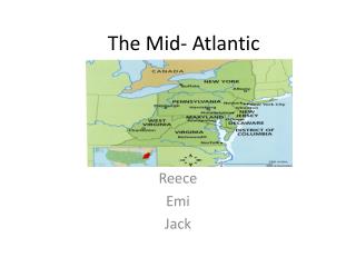
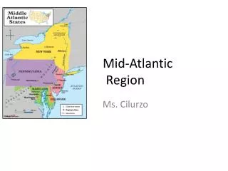
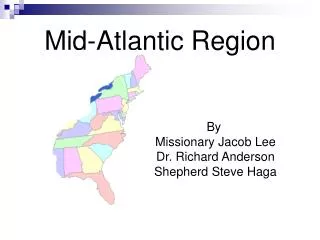
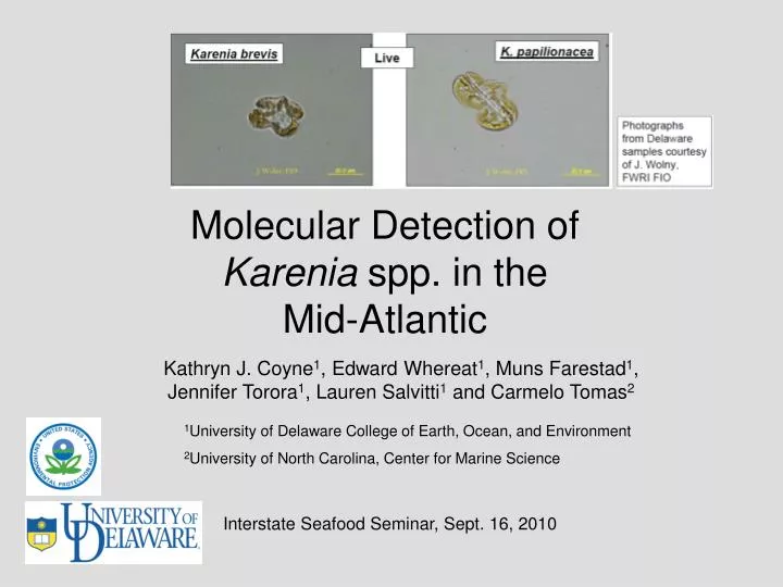
Molecular Detection of Karenia spp. in the Mid-Atlantic. Kathryn J. Coyne 1 , Edward Whereat 1 , Muns Farestad 1 , Jennifer Torora 1 , Lauren Salvitti 1 and Carmelo Tomas 2. 1 University of Delaware College of Earth, Ocean, and Environment

E N D
Molecular Detection of Karenia spp. in the Mid-Atlantic Kathryn J. Coyne1, Edward Whereat1, Muns Farestad1, Jennifer Torora1, Lauren Salvitti1 and Carmelo Tomas2 1University of Delaware College of Earth, Ocean, and Environment 2University of North Carolina, Center for Marine Science Interstate Seafood Seminar, Sept. 16, 2010
Overview • Molecular approach • Description • Rationale • Development • Validation • Karenia spp. in Mid-Atlantic coastal waters • Timeline • Molecular (presence/absence) results • Validation and results of quantitative approach for enumeration • Morphological variability • Distributions of Karenia spp. in DE and NJ
C T A T T C A C G T T T G G A A T T C C G T C T A T A T C A A T T T G G A A T T C C G T C T A T A T C A A G C T G G A A T T C C G T What are Molecular Methods?
Molecular Methods • Polymerase chain reaction (PCR) • Specific amplification of DNA fragment from a specific gene DNA primers bracket the area to be amplified Polymerase enzyme copies DNA fragment bracketed by primers to produce a product of a single size.
Why do we use Molecular Methods for HAB Research? To determine presence/absence or abundance of HAB species Advantages over microscopy: • Increased sensitivity • Increased accuracy in detection • Applicable to mixed assemblages • Higher throughput
18S small subunit ITS1 5.8S ITS2 28S large subunit Species are identified and discriminated based on variations in DNA sequence. rRNA genes - 10s to 100s of copies Highly conserved regions -Group or genus-specific primers Variable regions -Species or strain-specific primers
Quantitative Real-Time PCR Ct Wide dynamic range:Linear over 8 orders of magnitude Sensitive: Detection of less than 4 copies of target DNA is possible
How do we validate PCR methods? STEP 1: Primer design STEP 2: QPCR assay development Design primers for target species or group Optimize primers for QPCR Evaluate sensitivity and range of detection Use primers to amplify DNA from several samples Determine accuracy Sequence DNA to make sure primers are specific No Yes QPCR results Cell counts Research and Monitoring Yes No
Karenia spp. in Mid-Atlantic 2007 August 30, 2007: Initial observation of K. papilionaceae in water samples. Aug. - Sept., 2007: Microscopic identification of K. pap in off-shore waters and ocean beaches. Sept. 6 - 7, 2007: Smaller “K. brevis -like” cells observed in water samples. 2008 June - Nov., 2008: Water samples (n=33) from 4 sites in DE processed for molecular analysis. 2009 Mar. - Nov., 2009: Water samples (n=30) from 4 sites in DE processed for molecular analysis. Aug. 26-27: Water samples (n=7) from NJ coastal bays processed for molecular analysis. 2010 March - present, 2010: Water samples from 4 sites in DE processed for molecular analysis.
Primer design and evaluation (Kpap) • Primers for K. pap were initially provided by Miguel de Salas (UTasmania) • Targets the 28S rRNA gene. • DNA was amplified by PCR from environmental samples collected Aug.-Sept., 2007. • Sequenced PCR products from 3 env. samples collected on 2 different days. • Comparison of DE strain of Kpap with Kpap sequences in GenBank from Hong Kong, NZ and Australia • Results: Primers are specific to Kpap, but sequence is slightly different • Later confirmed by sequence analysis of pure culture of NZ and DE strains provided by Dr. Carm Tomas (UNC) M 1 2 3 N
When did K. papilionaceae first appear in DE coastal waters? • DNA from inlet and Fenwick beach water samples collected in June and July, 2007 were tested. • K. pap was not present on June 25, 2007. • Samples collected on July 11, 2007 through the end of August were positive. Results suggest that K. pap was present early in the summer
K. pap PCR Screening (presence/absence) for DE coastal waters 2008-2009 • 2008: 33 samples from 4 sites. • 13 samples collected before Aug. 14th were negative for K. papilionacea • All 20 samples collected between Aug. 14th and Nov. 13th were positive for K. pap. • 2009: 33 samples from 4 sites. • 16 samples collected before Aug. 17th were negative for K. papilionacea • 1 sample collected on Aug. 3rd was positive (IR39) • All 14 samples collected between Aug. 17th and Nov. 4th were positive for K. papilionacea • 2010: Samples collected Mar. through present • Samples collected before Aug. were negative for K. pap
QPCR assay for K. papilionaceae New primers and QPCR probes were designed based on sequence data - Specific to NZ and DE strains DE isolate genomic DNA DE isolate positive control NZ isolate positive control NZ isolate positive control DE isolate pos cont and genomic DNA
QPCR Results for K. papilionaceae 2008 Samples Calibrator sample Cell Count Detection limit = 167 cells/L • QPCR results are comparable to cell counts when above limit of detection • QPCR is more sensitive than cell counts
Distribution of K. pap north of Delaware Samples collected along NJ on Aug. 26-27, 2009 K. pap was present from Cape May to Ocean City M 1 2 3 4 5 6 7 P N
Molecular detection of K. brevis • Culture obtained from CCMP to use as positive control • 2 Different primer sets designed targeting 28S rRNA gene • Primers for K. brevis RUBISCO (Gray et al. 2003) • PCR optimized using DNA from K. brevis culture Results: Environmental samples do not amplify with the 3 different K. brevis primer sets - Weak, non-specific amplification M 1 2 3 N 1 2 3 N Kpap Kbrev
Morphological Variability • Unialgal cultures of the DE strain of K. papilionaceae were established by Dr. Carm Tomas. • Small “K. brevis –like” cells were observed in these cultures. • Small and large cells were picked into separate tubes and sent for DNA analysis. Results: Both large and small cells amplified with K. pap primers but not with K. brevis primers. It’s possible that the “K. brevis –like” cells in environmental water samples were actually K. papilionaceae. Another possibility is that the “K. brevis –like” cells in environmental water samples were an unknown Karenia strain or species. More work needs to be done
Summary and Implications • K. papilionacea has been detected in DE coastal waters each year since 2007 • Endemic population or imported each year? • K. brevis-like cells have been observed but have not been detected using molecular methods • Morphological variability of K. papilionaceae or new strain/species of Karenia?
Questions? Funding provided by DNREC EPA Beach Grant