Download
1 / 100
1.1k likes | 1.5k Views
Data Acquisition, Representation and Reconstruction of medical images. Application of Advanced Spectral Methods. Acquisition Methods for medical images. X-Rays Computer Tomography (CT or CAT) MRI (or NMR)

E N D
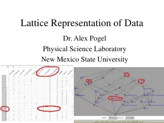
Data Acquisition, Representation and Reconstruction of medical images Application of Advanced Spectral Methods
Acquisition Methods for medical images • X-Rays • Computer Tomography (CT or CAT) • MRI (or NMR) • PET / SPECT (Positron Emission Tomography, Single Photon Emission Computerized Tomography • Ultrasound • Computational
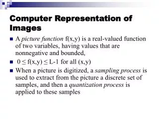
X-Rays - Physics • X-Rays are associated with inner shell electrons • As the electrons decelerate in the target through interaction, they emit electromagnetic radiation in the form of x-rays. • patient is located between an x-ray source and a film -> radiograph • cheap and relatively easy to use • potentially damaging to biological tissue
X-Rays • X-Rays • similar to visible light, but higher energy!
X-Rays - Visibility • bones contain heavy atoms -> with many electrons, which act as an absorber of x-rays • commonly used to image gross bone structure and lungs • excellent for detecting foreign metal objects • main disadvantage -> lack of anatomical structure • all other tissue has very similar absorption coefficient for x-rays
X-Rays - Images X-Rays can be used in computerized tomography
Non-Intrusive Medical Diagnosis based on Computerized Tomography • Computer tomography CT (From Jain’s Fig.10.1) An X-ray CT scanning system
Non-Intrusive Medical Diagnosis based on Transmission Tomography Source and Detector are rotating around human’s body (From Bovik’s Handbook Fig.10.2.1)
Non-Intrusive Medical Diagnosis based on projections Observe a set of projections (integrations) along different angles of a cross-section Each projection itself loses the resolution of inner structure Types of measurements transmission (X-ray), emission, magnetic resonance (MRI) Want to recover inner structurefrom the projections “Computerized Tomography” (CT)
Non-Intrusive Medical Diagnosis based on Emission Tomography • Emission tomography: ET measure emitted gamma rays by the decay of isotopes from radioactive nuclei of certain chemical compounds affixed to body parts. • MRI: based on that protons possess a magnetic moment and spin. • In magnetic field => align to parallel or antiparallel. • Apply RF => align to antiparallel. Remove RF => absorbed energy is remitted and detected by Rfdetector. f(x,y) is 2D image as before
Radon Transform Principles A linear transform f(x,y) g(s,) Line integral or “ray-sum” Along a line inclined at angle from y-axis and s away from origin Fix to get a 1-D signal g(s) We have now a set of images g(s) which represent g(s,) (From Jain’s Fig.10.2) This is a transform from 2D to 2D spaces
Tomography and Reconstruction Lecture Overview Applications Background/history of tomography Radon Transform Fourier Slice Theorem Filtered Back Projection Algebraic techniques • Measurement of Projection data • Example of flame tomography
Applications & Types of Tomography MRI and PET showing lesions in the brain. PET scan on the brain showing Parkinson’s Disease
Applications & Types of Tomography – non medical ECT on industrial pipe flows
CT or CAT - Principles Computerized (Axial) Tomography introduced in 1972 by Hounsfield and Cormack natural progression from X-rays based on the principle that a three-dimensional object can be reconstructed from its two dimensional projections based on the Radon transform (a map from an n-dimensional space to an (n-1)-dimensional space) Radon again! From 2D to 3D !
CT or CAT - Methods measures the attenuation of X-raysfrom many different angles a computer reconstructs the organ under study in a series of cross sections or planes combine X-ray pictures from various angles to reconstruct 3D structures
The History of CAT • Johan Radon (1917) showed how a reconstruction from projections was possible. • Cormack (1963,1964) introduced Fourier transforms into the reconstruction algorithms. • Hounsfield (1972) invented the X-ray Computer scanner for medical work, (which Cormack and Hounsfield shared a Nobel prize). • EMI Ltd (1971) announced development of the EMI scanner which combined X-ray measurements and sophisticated algorithms solved by digital computers.
Backpropagation Principles
Backpropagation We know that objects are somewhere here in black stripes, but where?
Example of Simple Backprojection Reconstruction Given are sums, we have to reconstruct values of pixels A, B, C and D
Image Reconstruction: ART or Algebraic Reconstruction Technique ART
CT - Reconstruction: ART or Algebraic Reconstruction Technique METHOD 1: Algebraic Reconstruction Technique iterative technique attributed to Gordon Initial Guess Reconstructedmodel Back-Projection Projection Actual DataSlices
CT - Reconstruction: FBP Filtered Back Propagation METHOD 2 : Filtered Back Projection common method uses Radon transformandFourier Slice Theorem y F(u,v) f(x,y) Gf(r) x s u gf(s) f Spatial Domain Frequency Domain
COMPARISON : CT - FBP vs. ART Computationally cheap Clinically usually 500 projections per slice problematic for noisy projections ART FBP Algebraic Reconstruction Technique • Still slow • better quality for fewer projections • better quality for non-uniform project. • “guided” reconstruct. (initial guess!) Filtered Back Projection
Fourier Slice Theorem and FFT review Patient’s body is described by spatial distribution of attenuation coefficient
Properties of attenuation coefficient Our transform: f(x,y) p(r,)
attenuation coefficient is used in CT_number of various tissues These numbers are represented in HU = Hounsfield Units CT_number uses attenuation coefficients
RADON TRANSFORM Properties REMEMBER: f(x,y) p(r,)
Radon Transform is available in Matlab • Radon and its inverse easy to use • You can do your own projects with CT reconstruction • Data are available on internet sinogram
Inverse Radon Transform Inverse Radon Transform
In Matlab there are implemented functions that use Fourier Slice Theorem Matlab example
Matlab example: filtering High frequency removed Low frequency removed
Remainder of main theorem of spectral imaging Matlab example – filtering by convolution in spectral domain
Example of Image Radon Transform [Y-axis] distance, [X-axis] angle (From Matlab Image Processing Toolbox Documentation)
big noise No noise