Download
1 / 41
430 likes | 858 Views
Pneumonia 101. Armaan Khalid. What the. Definition of Pneumonia. An acute or chronic disease marked by inflammation of the lung parenchyma, that causes consolidation of inflammatory exudates Main causes Bacteria Virus Fungal & etc. Classification. Anatomical/Radiological Lobar

E N D
Pneumonia 101 Armaan Khalid
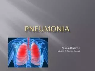
Definition of Pneumonia • An acute or chronic disease marked by inflammation of the lung parenchyma, that causes consolidation of inflammatory exudates • Main causes • Bacteria • Virus • Fungal & etc
Classification • Anatomical/Radiological • Lobar • Multi-focal/lobular (bronchopneumonia) • Interstitial (focal diffuse) • Location of Contraction • Community • Institutional (nursing home) • Nosocomial (hospital)
Precipitating Factors • Smoking (Smokers in household) • Previous lung pathology (COPD, CF) • EToH abuse • Immunosuppresion • Recent hospital admission • IVDU (S Aureus haematogenous spread) • Recent exposure to pneumonia pts • Preceding viral infection • HIV
Atypical Pneumonia • Assoc w a milder form of pneumonia • Walking pneumonia • Considered atypical because • Inability to detect on gram stain • Inability to be cultivated in normal media • Examples • Mycoplasma • Chlamydophila species • Legionella • Coxiella burnetii (Q fever) • Bordetella pertussis (Whooping cough)
Clinical Presentation • Preceding Hx of viral illness • On Hx/Ex • Febrile/Pleuritic Pain/Dry cough • Sputum production • Malaise/Rigors/Chills • Tachypnoea/cardia • ↓ chest movements • Use of accessory chest muscles • Sg of consolidation +/- pleural rub
History Taking • Impt to review pt’s: • Potential exposure • Envt/Work/Social factors • Aspiration risks • Seizure/EToH/GORD • Host factors • COPD/IVDU/Smoking/HIV
Sputum Characteristics • S Pneumoniae • Rust coloured sputum • Pseudomonas/Haemophilus & Pneumococcal • Green sputum • Klebsiella species • Red currant jelly sputum • Anaerobic species • Foul smelling/Bad tasting sputum
Risk Stratification • How do you make the decision to Rx the pt in a out/in-patient setting? • CURB-65 criteria • Pneumonia Severity Index (PSI) • PSI calculator online • http://pda.ahrq.gov/clinic/psi/psicalc.asp
CURB-65 criteria • C – Confusion • U – Uraemia, BUN > 20 mg/dL • R – Respiratory Rate > 30 bpm • B – Blood pressure < 90/60 mm Hg • 65 – Age > 65 years old • Score 0-1: Outpatient treatment • Score 2: Admit to the wards • Score 3-4: Admit to ICU
Differential Diagnosis • Asthma • Atelectasis • Bronchiectasis • COPD • Lung Abscess • Viral infection • Influenza
Workup • FBE/UNE/BUN/LFT/CRP/ESR • Blood cultures • Impt to get them before initiating empirical therapy • Sputum (microscopy & culture) • ABG • ? Pleural fluid tap • CXR (frontal & lateral)
Further Workup • Pneumococcal antigen • Counter-immunoelectrophoresis of sputum, urine & serum • Mycoplasma antibodies • Legionella & Chlamydia antibodies • Immunoflurorescent tests • Legionella antigen • Urinary antigen test
Radiological Findings • General Characteristics • Affected tissue will appear denser • May contain air bronchogram(s) • Visibility of air in the bronchi • Sign of airway disease, not pathognomonic for pneumonia • Airspace pneumonia appears fluffy & their margins are indistinct • If it abuts a pleural surface, there will be a sharp demarcation of the margins
Patterns of Appearance • Lobar • Segmental (Bronchopneumonia) • Interstitial • Round • Cavitary
Patterns on CXR • Lobar Pneumonia • Common organism: S Pneumoniae • Homogenous consolidation w air bronchogram • Silhouette sign present when in contact with the heart, aorta or diaphragm
Patterns on CXR • Segmental (Bronchopneumonia) • Common organisms: S Aureus & gram-negative bacteria • Affects the walls of the bronchioles • Spread centrifugally via tracheobronchial tree to many foci @ the same time • Margins are fluffy & indistinct • Produces exudate that fills the bronchi • No air bronchograms present • May be assoc w atelectasis
Patterns on CXR • Interstitial Pneumonia • Common organisms: Mycoplasma, viral pneumonia & PCP • Reticular interstitial disease w diffuse spread throughout lungs in early disease process • Frequently progresses to airspace disease
Patterns on CXR • Round Pneumonia • Common organisms: H influenzae, Strep & Pneumococcus • Spherical pneumonia usually seen in the lower lobes of children • May resemble a mass • Clinical presentation does not match w that of a mass
Patterns on CXR • Cavitary Pneumonia • Common organism: M tuberculosis • Primary TB < Reactivation TB • Primary TB • Upper lobes > lower lobes • Assoc w ipsilateral hilar adenopathy & large unilateral pleural effusions • Reactivation TB • Cavities are thin-walled, smooth inner margin & usually no air-fluid level
Spine Sign • On Lateral CXR, thoracic spine vertebra are darker in diaphragm than in shoulder girdle • CXR needs to penetrate more tissue in the shoulder girdle than in diaphragm • With interstitial/airspace disease in posterior lower lobe, vertebra would be more opaque (brighter) than usual • Spine Sign!
Silhouette Sign • If 2 objects of the same radiographic density touch each other, then their edges disappear • Silhouette Sign • Valuable in localising lung pathology
Management • Respiratory Support • O2 +/- bronchodilators • Fluid resuscitation • Empiric Abx Rx • Empiric Rx should initially be broad • Each hospital has it’s own guidelines
Supportive Measures • Analgesia & anti-pyretics • Chest physiotherapy • IV fluids or diuretics • Positioning of patient (Aspiration risk) • Suctioning & bronchial hygiene
Clinical Resolution • Clinical response to Abx Rx • Improvement seen in 48-72 hrs • Abx shouldn’t be changed w/in 72hrs • Time required for Abx to act • Change if marked deterioration • Radiological resolution takes longer than clinical resolution
Clinical Resolution (or lack thereof) • No resolution • Resistant to Abx • 2° to complications (empyema/abscess) • Non-infectious cause (CHF/malignancy) • Viral aetiology • Consider • CT/MRI • Bronchoscopy • Lung biopsy • Consult ID physician
Viral Pneumonia • Common in children & the elderly • Prevalent in the immunosuppressed • Uncommon in adults • 13-50% of all CAP • Influenza virus main offender (>50%) • Clinical findings similar to bacteria • May predispose & superimpose on a bacterial pneumonia • Common during winter • Rx • Supportive Rx • Antiviral • Immunisations
References • Kumar & Clark, Clinical Medicine, 6th edn, Chapter 14, Pneumonia, pp 922-929 • W Herring, Learning Radiology: Recognizing The Basics, 1st edn, Chapter 8 Recognizing Pneumonia, pp 60-67 • Longmore et al, OHCM, 7th edn , Chapter 5, Chest Medicine, pp 152-153