Download
1 / 45
450 likes | 592 Views
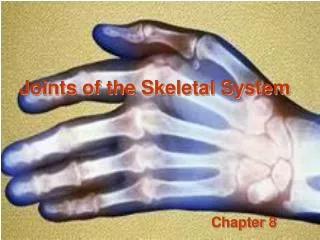
Joints of the Skeletal System . Advanced Biology Fall 2012 . Vocab Development . Anul - ring Arth - joint Burs- bag, purse Glen- joint socket Labr - lip Ov - egg-like Sutur - sewing Syn - with, together Syndesm - band, ligament . Introduction .

E N D
Joints of the Skeletal System Advanced Biology Fall 2012
Vocab Development • Anul- ring • Arth- joint • Burs- bag, purse • Glen- joint socket • Labr- lip • Ov- egg-like • Sutur- sewing • Syn- with, together • Syndesm- band, ligament
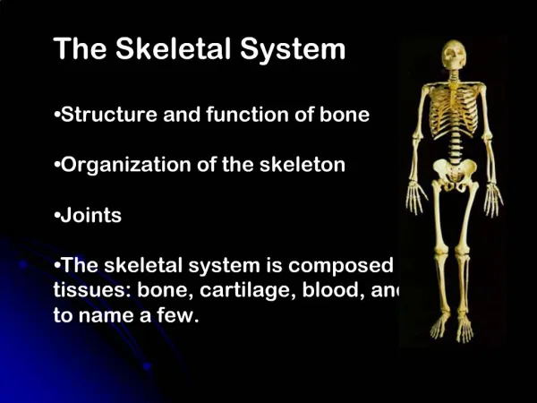
Introduction • Joints or articulations- functional junctions between bones • Bind parts of the skeletal system • Allow for bone growth • Permit parts of the skeleton to change shape during childbirth • Enable the body to move in response to skeletal muscle contractions
Introduction cont… • Joints vary in structure and function • Classified both structurally and functionally • Structurally by the type of tissue that binds the bones at each junctions • Functionally by the degree of movement possible
Introduction cont… • Structural joint classifications • Fibrous • Cartilaginous • Synovial • Functional joint classifications • Immovable (synarthrotic) • Slightly movable (amphiarthrotic) • Freely movable (diarthrotic)
3 types of Fibrous Joints • Syndesmosis • bones are bound by a sheet or bundle of dense connective tissue • Flexible & may be twisted • Permits slight movement (amphiarthrotic) • Ex: lies between the tibia & fibula
3 types of Fibrous Joints • Sutures • Only between the flat bones of the skull • Bones are united by a thin layer of dense connective tissue • Immovable (synarthrotic)
3 types of Fibrous Joints • Gomphosis • Formed by the union of a cone-shaped process in a bony socket • Ligament surrounds the root & firmly attaches it • Synarthrotic • Ex: teeth
2 types of Cartilaginous Joints • Synchondrosis • Bands of hyaline cartilage unite the bones • Many are temporary & disappear during growth • Synarthrotic • Ex: epiphyseal plates • Ex: between manubrium & first rib
2 types of Cartilaginous Joints • Symphysis • Covered by a thin layer of hyaline cartilage & the cartilage is attached to a pad of fibrocartilage • Limited movement • Ex: pubic symphysis • Ex: intervertebral discs
Synovial Joints • Most joints of the skeleton are synovial • Allow free movement (diarthrotic) • Consist of: articular cartilage, a joint capsule, & a synovial membrane
Synovial Joints • General Structure • Articular cartilage- covers the ends of the bones • Joint capsule- has 2 layers that hold together the bones of the joint • Synovial membrane-covers all surfaces with in the joint capsule (only a few cell layers thick) • synovial fluid- lubricates articular surfaces • Menisci- divide the synovial joints into some small compartments
Synovial Joints • General Structure cont… • purpose of the menisci is to help cushion the joint • Bursae (bursa sacs)- found in certain types of synovial joints • Help cushion and provide protection for bones that are close to the skins’ surface • Helps ligaments and tendons glide smoothly over bones
Synovial Joints • 6 types of synovial joints • Ball-and-socket joints- hip joint & shoulder joint • Condylar joint- where your finger bend (in between the phalanges) • Plane joints (gliding joints)- found in between your carpals & tarsals • Hinge joints- elbow & knee • Pivot joint- between the axis and atlas, between your radius and ulna • Saddle joint- where the thumb joins the carpals
Types of Joint Movements • Flexion- bending parts of a joint so that the angle between them decreases and the parts come closer together • Ex: bending knee • Extension- moving parts of a joint so that the angle between them increases and the parts move farther apart • Ex: straightening the knee
Types of Joint Movements • Hyperextension- used to describe the extension of the parts at a joint beyond the normal range of motion (usually causes injury) • Dorsiflexion- movement at the ankle that brings the foot closer to the shin • Ex: rocking back on your heels • Plantar Flexion- movement at the ankle that brings the foot farther from the shin • Ex: standing on your toes or walking
Types of Movements • Abduction- moving a part away from the midline or the axial line of the limb • Ex: spreading your fingers or toes • Adduction- moving a part toward the midline or toward the axial line of the limb • Ex: moving your fingers or toes closer together
Types of Movements • Rotation- moving a part around an axis • Medial rotation- turning of a limb toward the midline • Lateral rotation- turning of a limb away from the midline • Circumduction- moving part so that its end follows a circular path • Ex: moving a finger in a circular motion without moving your hand
Types of Movements • Supination- rotation of the forearm so the palm is upward • Pronation- rotation of the forearm so the palm is down • Eversion- turning the foot so the plantar surface faces laterally • Ex: big toe pushed down, little toe up • Inversion- turning the foot so the plantar surface faces medially • Ex: big toe up, little toe down
Types of Movements • Protraction- moving a part forward • Retraction- moving a part backward • Elevation- raising a part • Depression- lowering a part
Shoulder Joint • Ball and socket joint • Consists of the humerus & glenoid cavity • Ligaments • Coracohumeral ligaments • Connects the coracoid process of the scapula to the humerus • Glenohumeral ligaments (3) • Connect the glenoid cavity to the neck of the humerus
Ligaments… • Transverse Humeral Ligament • Across the humerus • several bursae • Labrum • Can perform: flexion, extension, adduction, abduction, rotation, and circumduction
Elbow Joint • 2 articulations • Hinge joint between the humerus & ulna • Planar joint between the ulna and humerus (only when elbow is bent, not when in anatomic position) • Ligaments • Ulnar collateral ligament • Connects the humerus to the ulna • Radial collateral ligament • Connects the humerus to the radius • Anular ligament • Connects the ulna to the radius at the head of the radius • Movements: flexion & extension • Supination and pronation of forearm is allowed by the anular ligament
Hip Joint • Ball and socket joint • Consists of: • Head of the femur • Acetebulum of the hip bone • Major Ligaments • Iliofemoral ligament • Strongest ligament in the body • Connects the iliac spine to the femur • Pubofemoral ligament • Runs from the superior pubis to the iliofemoral ligament • Ischiofemoral ligament • Runs from the ischium to the joint capsule
Hip Joint • Movements: • Flexion, extension, adduction, abduction, rotation, and circumduction
Knee Joint • Largest and most complex synovial joint • Consists • Femur • Tibia • Patella • Lots of ligaments
Knee Joint • Ligaments (these strengthen the joint capsule) • Patellar ligament • Extends from the patella to the tibia • Oblique popliteal • Connects the lateral condyle of the femur to the head of the tibia • Arcuatepopliteal • Extends from the lateral condyle of the femur to the head of the fibula • Tibial collateral (MCL) • Connects the medial condyle of the femur to the medial condyle of the tibia • Fibular Collateral (LCL) • Connects the lateral condyle of the femur and the head of the fibula
Knee Joint • Cruciate ligaments • Help prevent displacement of the articulating surfaces • Stretch upward and cross between the tibia and femur • Anterior cruciate ligament (ACL) • Runs from the anterior side of the tibia to the lateral condyle of the femur • Posterior cruciate ligament (PCL) • Connects the posterior side of the tibia to the medial condyle of the femur
Knee Joint • 2 menisci • Separate the articulating surfaces of the femur and tibia & help keep them aligned • Several bursae • Movement: flexion, extension, and some rotation
Joint Disorders • Sprains • Overstretching or tearing of connective tissues associated with a joint • Painful & swollen, restricts movement • Bursitis • Inflammation of the bursa • Caused by overuse or a sudden increase in activity • Ex: tennis elbow
Joint Disorders cont… • Arthritis • Causes inflamed, swollen, and painful joints • More than 100 different types • Can be part of other syndromes • Most common types are: rheumatoid , osteo, Lyme
Joint Disorders cont… • Rheumatoid Arthritis (RA) • Autoimmune condition • Painful & debilitating • Synovial membrane becomes hard and forms a mass • Over time joints can ossify • Usually a systemic illness
Joint Disorders cont… • Osteoarthritis • Degenerative disorder • Most common type of arthritis • Usually occurs with aging • Articular cartilage disappears slowly • Painful & restricts movement • NSAIDS are used to treat it along with regular exercise
Joint Disorders cont… • Lyme Arthritis • Caused by lyme disease and is intermittent • Antibiotic treatments early on can help treat it • Arthritis may also be associated with other bacterias like strep, staph, gonorrhea, tuberculosis • AIDS may also be associated with arthritis because it is an autoimmune disorder
Joint Disorders cont… • Other types of arthritis • Gout • Juvenile RA • Scleroderma • Systemic lupus erythemoatosus • Kawasaki disease • Strep A infection
http://youtu.be/HxJO8GaoZrk shoulder • http://youtu.be/dZiDd6e4drcacl