Download
1 / 1
10 likes | 127 Views
Pichot et al Supplemental Figure 1. B. IC50=17.8 μM. MDA-231. MDA-468. SKBR-3. T47D. A. MDA-MB-468. MCF-7. Dasa. Doxo. Combo (nM). DMSO. 100. 100. 100. 10. 10. 10. pSrc416. 1. pSrc-Y416 Src. c-kit. GAPDH. C. Supplemental Figure 1:

E N D
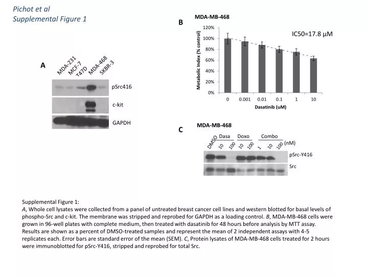
Pichot et alSupplemental Figure 1 B IC50=17.8 μM MDA-231 MDA-468 SKBR-3 T47D A MDA-MB-468 MCF-7 Dasa Doxo Combo (nM) DMSO 100 100 100 10 10 10 pSrc416 1 pSrc-Y416 Src c-kit GAPDH C Supplemental Figure 1: A, Whole cell lysates were collected from a panel of untreated breast cancer cell lines and western blotted for basal levels of phospho-Src and c-kit. The membrane was stripped and reprobed for GAPDH as a loading control. B, MDA-MB-468 cells were grown in 96-well plates with complete medium, then treated with dasatinib for 48 hours before analysis by MTT assay. Results are shown as a percent of DMSO-treated samples and represent the mean of 2 independent assays with 4-5 replicates each. Error bars are standard error of the mean (SEM). C, Protein lysates of MDA-MB-468 cells treated for 2 hours were immunoblotted for pSrc-Y416, stripped and reprobed for total Src.