Download
1 / 43
430 likes | 436 Views
Learn about the different organs and structures of the digestive system, their functions, and how they work together to break down and absorb food.

E N D
Definitions The Digestive System: Is the system of the body formed of all the organs responsible for the ingestion, digestion and absorption of food. It includes the alimentary canal and the accessory glands. The Alimentary Canal: Is a long continuous tube that extends from the mouth to the anus. It includes: the mouth, parts of the pharynx, the esophagus, the stomach, the small intestine and the large intestine. The Gastrointestinal Tract (GIT) includes the stomach and intestines only. The Accessory Glands These produce various secretions that help in the chemical breakdown of food. They include the salivary glands, the liver and pancreas. Food, however, does not pass through them.
Wall of the Gastrointestinal Tract • Wall of the alimentary canal from esophagus to anal canal has same basic 4 layers. • Some difference are present in the different organs. • Mucosa – inner lining • Epithelium - protection, secretion, absorption • Lamina propria – connective tissue with blood and lymphatic vessels and lymphatic tissue. • Muscularis mucosae – thin layer of smooth muscle. • Submucosa • Connective tissue binding mucosa to muscularis. • Contains many blood and lymphatic vessels. • Submucosal plexus of nerves.
Muscularis • Voluntary skeletal muscle found in mouth, pharynx, upper 2/3 of esophagus, and anal sphincter. Involuntary smooth muscle elsewhere. • Arranged in layers (usually 2). • Contains the myenteric plexus between muscle layers. • The submucosal and myenteric plexuses form the enteric nervous system. • Serosa • Outermost covering of organs suspended in abdominopelviccavity. Formed of connective tissue and epithelium. • Also called visceral peritoneum. • Parts of GI tract not covered by peritoneum lack serosa – has adventitia. Example: esophagus, parts of duodenum, ascending and descending colon. Adventitia is formed of connective tissue only
The Abdominal Cavity • Borders: • Anterior: The anterior abdominal wall. • Posterior: Posterior abdominal wall. • Superior: The diaphragm. • Inferior: The pelvic brim. • Below the pelvic brim the cavity called the pelvic cavity. The abdominal and pelvic cavities are continuous with each and together called the abdominopelvic cavity.
Body cavities: Fig.3*: Lateral view of the body showing body cavities.
Peritoneum • Largest serous membrane of the body. • Divided into: • Parietal peritoneum – lines wall of cavity. • Visceral peritoneum – covers some organs ( = Serosa). • Space between is peritoneal cavity and it contain the peritoneal fluid. • 5 major peritoneal folds: • Two layers of peritoneum connecting organs to each other and to the anterior and posterior abdominal walls. • Greater omentum, lesser omentum, falciformligament, mesentery, and mesocolon • The space between the two walls of the greater omentum is called the lesser sac (located behind the stomach). The rest of the peritoneal cavity is called the greater sac.
Lesser sac Greater sac Fig.4: The peritoneum and its folds.
The Mouth (Oral/Buccal Cavity) • Bordered by the cheeks, lips, hard and soft palates, and the tongue. • Vestibule of the mouthis the space between the cheeks and lips externally and the gum and teeth internally. • Oral cavity properis the space that extends from the gums and teeth to the fauces. • The cheeks and lips help keep the food in the mouth. Fig.6: The mouth and its borders. • The mouth contains the tongue, the teeth and gums and into it open the ducts of salivary glands.
The Tongue • Skeletal muscles covered by mucous membrane. • Maneuvers food for chewing and forces food back for swallowing. • Contains Lingual glands which secrete lipase. • Dorsal surface has 3 types of projections called lingual papillae: Filiform, Fungiform Fig.8: The tongue. • and Vallate. The vallate type is the least numerous but it’s the largest and contains most of the taste buds (for the sense of taste). Fungiform papillae also has taste buds. Filiform papillae has no taste buds but assist in moving food around the mouth.
The Teeth: (Fig.9*) • Covers Dentin of crown • Hardest substance in the body 1 Above the gum 2 Blood and lymphatic vessels and nerves Within the gum 3 Within the bone Attaches root of teeth to bone Function: break down food into smaller particles (mastication)
The Salivary Glands • Salivary glands release saliva into the mouth: • Ordinarily, just enough is secreted to keep mouth and pharynx moist and clean. • When food enters mouth, secretion increases to lubricate mouth, dissolve food and begin chemical digestion. • 3 pairs of major salivary glands secrete most of the saliva: • Parotid: The largest. Located anterior and inferior to the ear. Its duct opens in the upper part of the oral cavity. • Submandibular: Located near the angle of the mandible. Its duct opens in the floor of the mouth. • Sublingual: Located under the tongue. Its ducts open in the floor of the mouth.
The Esophagus • A long muscular tube • Course: • Begins in the neck at the level of C6 vertebra. • Runs down posterior to the trachea. • Enters the superior mediastinum and then passes into the posterior mediastinum where it runs just anterior to the vertebral column. • Passes through the esophageal hiatus of the diaphragm (T10 vertebra) to enter the abdomen. • Enters the stomach at the cardia. • It’s closely related to the trachea, vertebral column, pericardium and the left atrium, descending thoracic aorta and the diaphragm.
Themuscularis layer of the wall of the esophagus is formed of: • Upper part – Skeletal muscles • Middle part – Skeletal and smooth muscles • Lower part – Smooth muscles • The esophagus is a passageway for the food from the pharynx to the stomach and it, thus,plays a part in deglutition • To facilitate this, the esophagus secretes some mucous. And it pushes food down by a series of muscle contractions and relaxations called peristalsis. Fig.12*: The process of swallowing.
The Stomach • A J-shaped dilation of the alimentary tract located inferior to the diaphragm in the left hypochondriac and epigastric regions and even the umbilical region when it’s distended. • The stomach churns and breaks down food and it mixes the food with its digestive secretions converting it into the semisolid chyme. The stomach also acts as a reservoir for the food. • Mucosa – gastric glands: • 3 types of exocrine gland cells – mucous cells (mucus), parietal cells (HCl and intrinsic factor), and chief cells (pepsinogen and gastric lipase). • G cell – endocrine cell – secretes gastrin. • The muscularis of the stomach is formed of 3 layers.
Parietal Cells: • Pyramidal in shape • Central nucleus • Abundant mitochondria • Special features depends on activity Fig.14*: Parietal cell. • In the resting phase, the canaliculus is short and the microvilli are absent. The cytoplasm is filled with tubulo-vesicles that fuse with the cell membrane when the cell is activated to produce the deep canaliculus and the microvilli. In the active phase, the cell has a deep intracellular canaliculus, into which protrude numerous microvilli. This increases the surface area for secretion. Fig.15*: Parietal cell features when active and resting.
Anatomy of the stomach • The stomach can be divided into 4 regions: • The Cardia – the part that surrounds the opening of the esophagus. Fig.16: Anatomical features of the stomach. • The Fundus – located above and to the left of the cardia. Usually filled with gas. • The Body – The main part of the stomach. • The Pyloric region – The distal part of the stomach formed of the distended pyloric antrum, the pyloric canal and pylorus. The pylorus opens into the duodenum and possesses the pyloric sphincter (a thickening in the muscularis layer).
The longer left border of the stomach is called the greater curvature. The shorter right border is called the lesser curvature. • At the junction between the esophagus and the stomach, there’s an angle called the cardiac angle. • The wall of an empty stomach shows several folds called rugae. These increase surface area. • The stomach is related to the lesser sac spleen, pancreas, left suprarenal gland, left kidney, the splenic artery, the transverse colon and the diaphragm.
The Small Intestine • It’s a long coiled tube formed of 3 parts – duodenum, jejunum, and ileum. • Its function is the digestion of food particles delivered from the stomach and the subsequent absorption of nutrients. • Its wall is characterized by the presence of folds called the plicae circulares. • The submucosa of the duodenum features numerous mucous glands. The submucosa of the ileum features numerous large lymphatic nodules called Payer's patches.
The duodenum is a C-shaped tube connecting the stomach with jejunum. • The concavity of the C is directed to the left and is occupied by the head of the pancreas. • The duodenum receives the duct of the pancreas and the bile duct. Fig.18: The small intestine. • The jejunum is located mostly in the left upper quadrant of the abdomen. Most absorption takes place here. • The ileum is located mostly in the right lower quadrant. It opens into the cecum. This opening is guarded by the ileocecal valve.
Features that increase the surface area of the small intestine: • Circular folds (Plicaecirculares) • Permanent ridges of mucosa and submucosa. • Cause chyme to spiral, delaying its passage allowing more time for absorption. • Villi • Fingerlike projections of mucosa • Contains arteriole, venule, blood capillary, and lacteal (lymphatic). • Microvilli • Projections of apical membrane of absorptive cells. • Contains digestive enzymes. Fig.19: Plicae and villi.
The Large Intestine • The terminal portion of the alimentary canal. • Overall function is to complete absorption (mainly water), produce certain vitamins (Vitamin K produced by intestinal bacteria), and form and expel undigested food (feces). • The longitudinal muscle layer of the muscularis is arranged in bundles called taenia coli. Because of their tone, they form pouches in the large intestine called haustra. • 4 major regions – cecum, colon, rectum, and anal canal. • The cecum is a blind pouch. To it is attached a blind tube called the appendix. The appendix contains several lymphatic nodules and, thus, plays an immune role. • Colondivided into ascending, transverse, descending and sigmoid colon. The colon is the part in which the main function of the large intestine takes place.
The rectum acts as a reservoir for the feces. It lies anterior to the sacrum and coccyx. • The anal canal is characterized by the presence of longitudinal folds called the anal columns. The opening of anal canal (anus) is guarded by the internal anal sphincter of smooth muscle and the external anal sphincter of skeletal muscle. Fig.21: The anal canal.
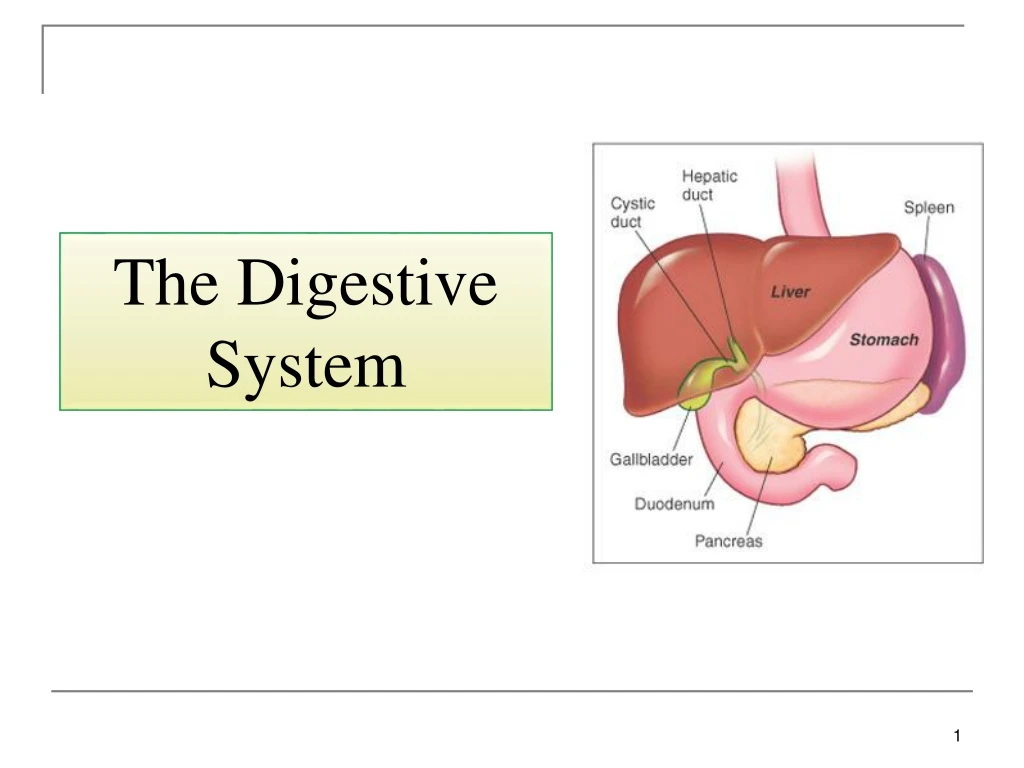
The Liver and Gallbladder • The liver is the largest internal organ and the heaviest gland in the body. • It’s located beneath the diaphragm in the right hypochondriac region and extends into the epigastric region. • Anatomically, it’s divided into right and left lobes by a fold of peritoneum called the falciform ligament. The right lobe also has the quadrate and caudate lobes. • Functions of the liver: • Metabolism of carbohydrates, lipids and proteins. • Detoxification. • Synthesis of bile and proteins. • Storage of glycogen, vitamins and minerals. • Activation of vitamin D. • Phagocytosis of RBCs.
Fig.22: The liver. The caudate and quadrate lobes are anatomically part of the right lobe, but functionally part of the left
Liver is composed of • Hepatocytes – major functional cells of liver with a variety of organelles: rough and smooth endoplasmic reticulum, Golgi apparatus, lysosomes and others. • Ito cells (stores vitamin A) and Kupffer cells (phagocytosis). • Bile canaliculi– narrow spaces between hepatocytes that collect bile secreted from the hepatocytes. Canaliculi unite to form small bile ducts. Eventually bile leaves the liver through the right and left hepatic ducts. These unite to form the common hepatic duct • Hepatic sinusoids – highly permeable blood capillaries receiving oxygenated blood from hepatic artery and deoxygenated nutrient-rich blood from hepatic portal vein. • The liver is surrounded by a capsule and the various components are arranged in the form of hexagonal hepatic lobules separated by connective tissue septa. At the center of these lobules is the central vein and at each corner we have a portal triad composed of: bile duct, branch of the hepatic artery and a branch of the portal vein all embedded in areolar connective tissue.
Liver receives blood from: Hepatic artery carrying oxygenated blood. Hepatic portal vein carrying deoxygenated blood with newly absorbed nutrients and possibly drugs, microbes or toxins from the alimentary tract. Blood flow through the liver
The Gallbladder • The gallbladder is a pear-shaped organ situated on the under surface of the liver. • It’s composed of 3 parts: a large fundus which protrudes from the anteroinferior margin of the liver, a body and a narrow neck (the last two are located under the liver). The neck opens into the cystic duct. The cystic duct joins the common hepatic duct to form the common bile duct. • Function: storage and concentration of bile and the release of bile into the small intestine when needed.
The Pancreas • The pancreas is an elongated gland located posterior to the stomach. It has both exocrine and endocrine function. • The pancreas is formed of 4 parts: the head, the neck, the body and the tail. • The head is the expanded right part of the pancreas. It’s located in the concavity of the duodenum. The head possesses a process called the uncinate process. • The neck is a constricted region after the head. To its left, the body passes upwards and to the left. The tail is the left tapering end of the pancreas that’s related to the spleen.
Histology • 99% of cells are acini: • Exocrine. • Secrete pancreatic juice – mixture of fluid and digestive enzymes. • 1% of cells are pancreatic islets (islets of Langerhans) • Endocrine. • Secrete hormones like glucagon, insulin and others. Fig.25: Histology of the pancreas.
Pancreatic juice secreted into the main and accessory pancreatic ducts and then pass to the small intestine: • The main pancreatic duct joins the common bile duct to form the hepatopancreaticampulla (of Vater). This opens into the duodenum at the major duodenal papilla which’s guarded by the sphincter of Oddi. • The accessory pancreatic duct opens into the minor duodenal papilla superior to the major duodenal papilla. Fig.26: The hepatopancreaticampulla.