Download
1 / 15
150 likes | 837 Views
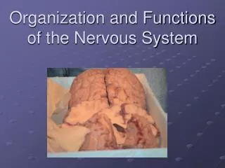
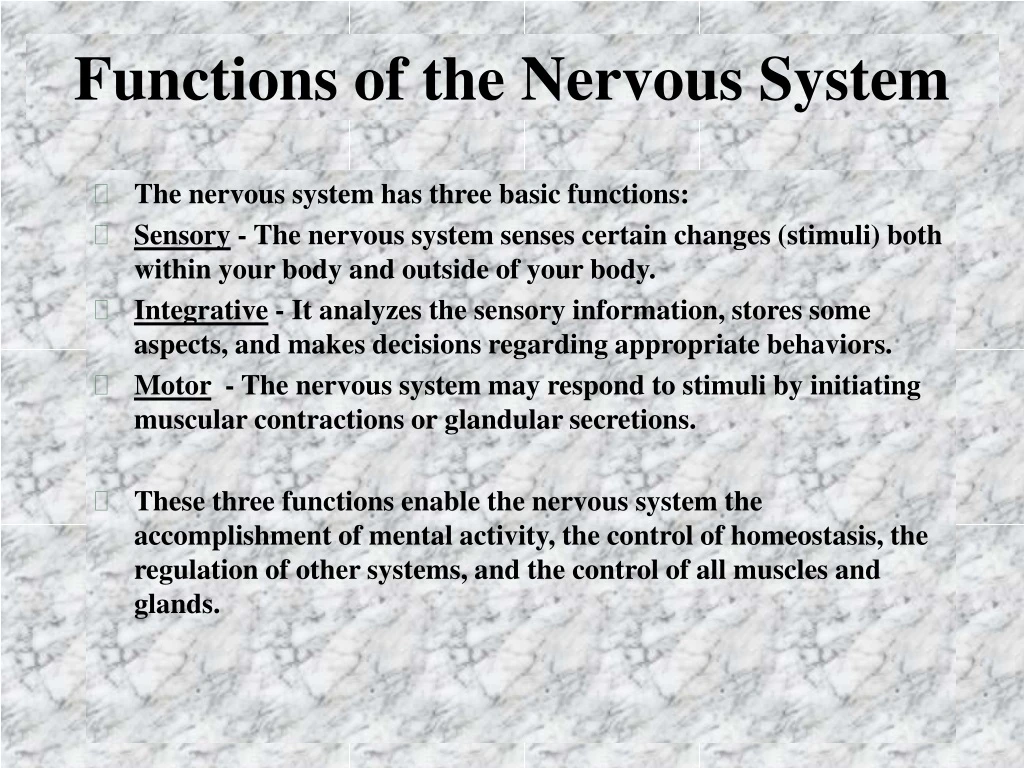
Functions of the Nervous System. The nervous system has three basic functions: Sensory - The nervous system senses certain changes (stimuli) both within your body and outside of your body.

E N D
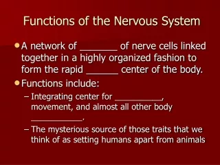
Functions of the Nervous System • The nervous system has three basic functions: • Sensory - The nervous system senses certain changes (stimuli) both within your body and outside of your body. • Integrative - It analyzes the sensory information, stores some aspects, and makes decisions regarding appropriate behaviors. • Motor - The nervous system may respond to stimuli by initiating muscular contractions or glandular secretions. • These three functions enable the nervous system the accomplishment of mental activity, the control of homeostasis, the regulation of other systems, and the control of all muscles and glands.
Divisions of the Nervous System • The central nervous system (CNS) consists of the brain and the spinal cord. • The peripheral nervous system (PNS) consists of nerves and ganglia outside the CNS. • The afferent division of the PNS transmits action potentials to the CNS. • The efferent division carries action potentials away from the CNS. • The PNS is subdivided into the somatic motor nervous system and the autonomic nervous system. • The somatic motor nervous system which innervates skeletal muscle is mostly under voluntary control. • The autonomic nervous system (ANS) innervates cardiac muscle, smooth muscle, and glands, and it is mostly under involuntary control. • The ANS has sympathetic and parasympathetic divisions.
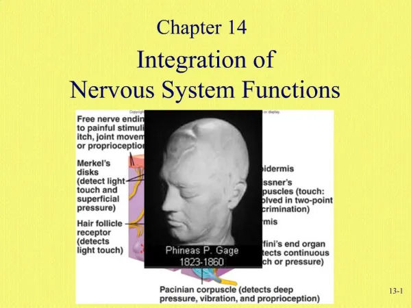
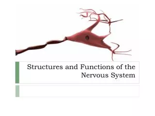
Histology of the Nervous System • The cells of the nervous system include neurons and neuroglia. • Neurons receive stimuli and transmit action potentials. • Neurons consist of a cell body (soma or parikaryon), dendrites, and axons. • Neurons can be multipolar, bipolar, or unipolar. • On the basis of function, sensory (afferent) neurons conduct impulses from receptors to the CNS; association neurons (interneurons) conduct impulses from one neuron to another within the CNS; and motor (efferent) neurons conduct impulses to effectors. • Neuroglia include astrocytes, oligodendrocytes, Schwann cells, microglia, and ependymal cells. • Neuroglia are specialized tissue cells that support neurons, attach neurons to blood vessels, produce myelin sheaths around axons, and carry out phagocytosis. • Two types of neuroglia produce myelin sheaths: oligodendrocytes myelinate axons in the CNS and Schwann cells myelinate axons in the PNS.
The Cerebellum • The cerebellum occupies the inferior and posterior aspects of the cranial cavity. It consists of two lateral hemispheres and a medial, constricted vermis. • The cerebellum is attached to the brain stem by three pairs of cerebellar peduncles. These peduncles contain both sensory and motor axons that carry information into and out of the cerebellum. • The cerebellum functions in the coordination of skeletal muscles and the maintenance of normal muscle tone, posture, and balance.
The Cerebrum • The cerebrum is the largest part of the brain. Its cortex contains gyri (convolutions), fissures, and sulci. • The cerebral lobes are named the frontal, parietal, temporal, and occipital. • The white matter is deep to the cortex and consists of myelinated and unmyelinated axons extending to other regions as association, commissural, and projection fibers. • The basal ganglia are paired masses of gray matter in the cerebral hemisphere. They help control muscular movements. • The limbic system is found in the cerebral hemispheres and diencephalon. It functions in emotional aspects of behavior and memory.
The Cerebrum • The sensory areas of the cerebral cortex are concerned with the interpretation of sensory impulses. • The motor areas are the regions that govern muscular movement. • The association areas are concerned with more complex integrative functions such as recognition of information. • Speech involves the sensory speech area, the motor speech area, and the interactions between them and other cortical areas. • Memory consists of sensory (lasting less than 1 sec), short-term (lasting a few minutes), and long-term (permanent) memory. • Each hemisphere controls the opposite half of the body. Commissures connect the two hemispheres. The left hemisphere is thought to dominate analytical, while the left is dominant for spatial perception and musical ability.
Neural Terms • Ganglia - A cluster of neuron cell bodies outside of the CNS. Located in the PNS. • Nucleus - A cluster of neuron cell bodies in the CNS. • Nerve - A group of axons and dendrites located in the PNS. • Fiber tract - A group of axons and dendrites located in the CNS. • Decussation- The crossing of nerve tracts from one side to the other within the CNS. • Contralateral - On the opposite side; affecting or controlling the opposite side of the body. • Ipsalateral - On the same side; affecting or controlling the same side of the body.
Development of the Nervous System • The development of the nervous system begins with a thickening of ectoderm called the neural plate. • The parts of the brain develop from primary and secondary vesicles. • The primary vesicles include the prosencephalon (forebrain), mesoencephalon (midbrain), and the rhombencephalon (hindbrain). • There are five secondary vesicles. • The prosencephalon divides into an anterior telencephalon and a posterior diencephalon. • The mesencephalon remains unchanged. • The rhombencephalon divides into an anterior metencephalon and a posterior myelencephalon.
Neural Crest Cell Derivatives • Bones of the skull • Pharyngeal arch cartilages • Otic capsule • Stroma of the thyroid, parathyroid, thymus and salivary glands. • Adrenal medullary cells • Enteric neurons • Sympathetic neurons • Sensory neurons • Schwann cells • Pigment cells