Download
1 / 70
700 likes | 728 Views
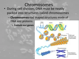
Chromosomes. Genetic material organization: condensation Nucleic acid: negative charged Proteins: positive charged Nucleoproteins: DNA(RNA)-protein complex. Bacteria: nucleoid Eukaryotic: 1-chromatin 2-chromosomes Packing ratio: DNA(RNA)-proteins. Nucleic Acid Molecules

E N D
Genetic material organization: condensation Nucleic acid: negative charged Proteins: positive charged Nucleoproteins:DNA(RNA)-protein complex Bacteria: nucleoid Eukaryotic: 1-chromatin 2-chromosomes Packing ratio: DNA(RNA)-proteins Nucleic Acid Molecules are longer than their compartments Figure 28.1 Packing ratio = the length of the DNA divided by the length of the unit that contains it.
Viral Genomes Are Packaged into Their Coats Tobacco mosaic virus • The length of DNA/RNA that can be incorporated into a virus is limited by the structure of the head-shell. How specific is the packaging of the viral genome? DNA(RNA) /protein Interaction Primary and tertiary structures Capsid (structural proteins and functional viral proteins) Nucleic acid (RNA) Protein shell is formed by a two- layer disk and each layer contains 17 identical proteins. RNA interacts with each layer of Proteins. Figure 28.2
Nucleic acid within the head-shell is extremely condensed (~4.8 kb) • Filamentous DNA viruses condense the DNA genome as they assemble the head-shell around it. • Spherical DNA viruses insert the DNA into a preassembled protein shell. Figure 28.3
Viruses use protein coats for packing Translocation (phage lambda) Cohesive Complementary ends 1-step:cleavage at the cos sequence 2-step: Binding of terminase (2 subunits) 3-step: recruitment to the head 4-step: translocation 5-step: condensation Head-shell (protein) and DNA. Insertion of DNA into the phage head. Translocation (ATP-dependent process) Condensation (mechanism: protein-scaffolding- ?) Cos sequences are ~200 base pairs long and essential for packaging Cos N site: cleavage site Cos B site: It holds the terminase Cos Q site: It protects from degradation (cellular DNAses)
The Bacterial Genome Is a Nucleoid • The bacterial nucleoid is ∼80% DNA by mass. • It can be unfolded by agents that act on RNA or protein. • The proteins that are responsible for condensing the DNA have not been identified. • HU and H1 proteins heat-unstable nucleoid protein Histone-like protein H1 (H-NS) High mobility group: Non-specific interaction Figure 28.5
The Bacterial Genome Is Supercoiled • The nucleoid has • ∼100 independent • negatively supercoiled • domains. • The average density • of supercoiling is • ∼1 supercoil/100 bp. Loops Organization of the genome into loops is associated with supercoiling of the DNA in vivo. Nicking the DNA relaxes the supercoils. Figure 28.7
Supercoiled DNA Supercoiling: DNA twisted around itself Positive supercoiled DNA: DNA is twisted around itself in the same sense as the two strands within the double helix (clockwise) Negative supercoiled DNA: DNA is twisted around itself in the opposite sense as the two strands within the double helix
DNA of interphase chromatin is negatively supercoiled into independent domains of ∼85 kb. • Metaphase chromosomes have a protein scaffold to which the loops of supercoiled DNA are attached. Eukaryotic interphase DNA is organized similarly to prokaryotic DNA. DNA loops are anchored in a protein scaffold that does not require DNA for formation. Eukaryotic DNA Has Loops and Domains Attached to a Scaffold DNA Protein Scaffold
Specific Sequences Attach DNA to an Interphase Matrix DNA is attached to the nuclear matrix at specific sequences called MARs (matrix associated regions) or SARs (scaffold attachment regions). The MARs are AT rich (>70%) but do not have any specific consensus sequence. S/MAR sequences may have role in regulating supercoiling of loops. . Figure 28.10
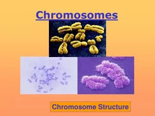
Chromatin is divided into euchromatin and heterochromatin Chromatin Is Divided into Euchromatin and Heterochromatin Chromosomes are compacted at Mitosis. Heterochromatin is more densely packed than euchromatin Heterochromatin : -permanently condensed -consists in DNA sequence repeats (not transcribed) -reduced density of genes (inactivated) -replicates at late states of the S phase. -interacts with Histones Individual chromosomes can be seen only during mitosis During interphase, the general mass of chromatin is in the form of euchromatin, which is less tightly packed than mitotic chromosomes. Regions of heterochromatin remain densely packed throughout interphase.
Chromosomes Have Banding Patterns • Certain staining techniques cause the chromosomes to have the appearance of a series of striations. • They are called G-bands. Figure 28.13 Giemsa staining allows visualization of chromosome binding patterns.
Arm//region//area/band=P22.1 Of 20kd fragment • The G bands are lower in G-C content than the interbands. • Genes are concentrated in the G-C-rich interbands. Certain staining techniques cause the chromosomes to have the appearance of a series of striations called G-bands G-(dark)bands are lower in GC content than interbands (high GC), genes are concentrated in the GC rich interbands (hybridization of mRNA).
DARK BANDS (G BANDS)PALE BANDS (CORRESPOND TO I BANDS) Stain strongly with dyes that bind preferentially to Stain weakly….. AT-rich regions such as Giemsa and Quinicrine May be comparatively AT-richMaybe comparatively GC-rich DNase insensitiveDNase sensitive Condense early during the cell cycle but replicate lateCondense late during cell cycle but replicate early Gene poor. Genes may be large because exons are often Gene rich, Genes are comparatively small because of close separated by very large introns clustering of exons LINE rich but may be poor in Alu repeatsLINE poor but may be enriched in Alu repeats
Special conditions for visualizing chromosomes and gene expression Visualization of condensed chromosomes that express genes during an extended meiotic period in some amphibians (lampbrush chromosomes). Visualization of amplified chromosomes (polytene chromosomes) in the larvae of Dipteran flies (Drosophila).
Lampbrush Chromosomes Are Extended • Sites of gene expression on lampbrush chromosomes show loops that are extended from the chromosomal axis. Figure 28.16 When are chiasmata formed? DNA recombination Why would the presence of ribonucleoprotein suggests gene expression? Nascent RNA chains
Polytene Chromosomes Form Bands • Polytene chromosomes of dipterans have a series of bands that can be used as a cytological map. (Drosophila) Dna probe (Labeled cDNA derived from a mRNA) Figure 28.18 Are these genes transcribed?
Polytene Chromosomes Expand at Sites of Gene Expression • Bands that are sites of gene expression on polytene chromosomes expand to give “puffs.” Characteristics of the puff material DNA is in more relax conformation Synthesis of RNA Transcription Proteins required for transcription (RNA polymerase II and others) Puff material site of transcription? Figure 28.21
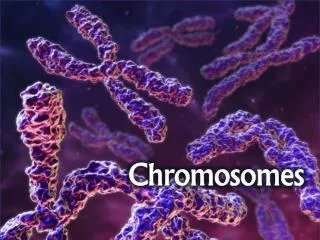
The Eukaryotic Chromosome Is a Segregation Device • A eukaryotic chromosome is held on the mitotic spindle by the attachment of microtubules to the kinetochore that forms in its centromeric region. • Centromeres often have heterochromatin that is rich in satellite DNA sequences ( highly repetitive DNA sequence). Figure 28.22
Centromeres are required for the correct segregation of chromosomes at mitosis and meiosis Centromeric DNA: Is formed by the heterochromatin DNA ( very common but not a 100 % of co-localization) C-bands (specific staining- DAPI or 4',6-diamidino-2-phenylindole, 33258 hoechst ) Conserved DNA sequence (responsible for DNA-protein [microtubules] interaction) Kinetochore- DNA and microtubules (part of the MTOC) Centromere or centromeric region: DNA: Proteins: Cohesins proteins (glue) Microtubules (MTOCs) Other proteins ? Centromere DNA sequences confer mitotic stability on pieces of dsDNA
Centromeres May Contain Repetitive DNA • Centromeres in higher eukaryotic chromosomes contain large amounts of repetitive DNA. • The function of the repetitive DNA is not known. Saccharomyces cerevisiae ~120 bp Schizosaccharomyces pombe ~40 to 100 kb Drosophila ~200 to 600 kb Arabidopsis >500 kb
Centromeres Have Short DNA Sequences in S. cerevisiae • Centromeric DNA regions (CEN) elements are identified in S. cerevisiae by the ability to allow a plasmid to segregate accurately at mitosis. • CEN elements consist of the short conserved sequences CDE-I and CDE-III. • They flank the A-T-rich region CDE-II. Figure 28.25 CDE: cycle-dependent element
The Centromere Binds a Protein Complex • A specialized protein complex that is an alternative to the usual chromatin structure is formed at CDE-II. • The CBF3 protein complex that binds to CDE-III is essential for centromeric function. • The proteins that connect these two complexes may provide the connection to microtubules. Mutations in CDE affect normal Segregation of DNA Figure 28.26
Telomeres Have Simple Repeating Sequences • The telomere is required for the stability of the chromosome end. • A telomere consists of a simple repeat where a C+A -rich strand has the sequence C>1(A/T)1–4. Figure 28.27
Telomeres Seal the Chromosome Ends • The protein TRF2 catalyzes a reaction in which the 3′ repeating unit of the G+T-rich strand forms a D-T loop (5 to 10 kb). • It displaces its homolog in an upstream region of the telomere. Figure 28.30
Telomeres Are Synthesized by a Ribonucleoprotein Enzyme • Telomerase (RT) uses the 3′–OH of the G+T telomeric strand to prime synthesis of tandem TTGGGG repeats. • RNA as template Figure 28.31
28.19 Telomeres Are Essential for Survival Figure 28.32
Why are telomeres important? The telomere can reach a length of 15,000 base pairs. Telomeres function by preventing chromosomes from losing base pair sequences at their ends. Telomeres also stop chromosomes from fusing to each other. However, each time a cell divides, some of the telomere is lost (usually 25-200 base pairs per division). When the telomere becomes too short, the chromosome reaches a "critical length”==>apoptosis. • Telomeres allow cells to distinguish chromosome ends from broken DNA ◦ If DNA is broken there are two options after the cell cycle is stopped: Repair or Death ▪ Repair can occur in two ways: ▪ Homologous Recombination (HR) -- ▪ Non-homologous end-joining (NHEJ) -- • Telomeres prevent chromosome fusions by NHEJ ◦ Fusion-bridge-breakage cycles leads to genomic instability which in turn can result in cell death or neoplastic transformation • Telomeres are specialized structures that are essential for protecting chromosome ends and ensuring chromosome stability
The Nucleosome Is the Subunit of All Chromatin • Micrococcal nuclease releases individual nucleosomes from chromatin as 11S particles. Endonucleases Figure 29.2
Nucleosomes are the fundamental subunits of chromatin in eukaryotes. The nucleosome is ~200 bp of DNA wrapped on the surface of a histone octamer (two H2AH2B dimers and one H3H4 tetramer). H1 is not required for the correct formation of the nucleosome, but is part of it. Nucleosome organization: Nucleosomes are the first level of organization (10 nm). Nucleosome fiber ( coiling of the series of nucleosomes) is the second level of organization (30 nm). The third level of organization is the packaging of the Nucleosome fiber itself. • A nucleosome contains “Basic structure “ • ∼200 bp of DNA • two copies of each core histone • (H2A, H2B, H3, and H4) Figure 29.3
DNA is wrapped around the outside surface of the protein octamer. Figure 29.5 ~80 bp of DNA makes two turn around the histones core (H1 is no included)
DNA Is Coiled in Arrays of Nucleosomes • >95% of the DNA is recovered in nucleosomes or multimers when micrococcal nuclease cleaves DNA of chromatin. • The length of DNA per nucleosome varies for individual tissues in a range from 154 to 260 bp. Chromatin (Nucleosomes)n Nuclease* *Experimental Conditions “Micrococcal” Buffer ~pH:8.0, 5mM CaCl2, 100mM NaCl. Figure 29.7/8
Nucleosomes Have a Common Structure • Nucleosomal DNA is divided into the core DNAandlinker DNA. • It depends on the DNA’s susceptibility to micrococcal nuclease. • The core DNA is the length of 146 bp that is found on the core particles produced by prolonged digestion with micrococcal nuclease. Nucleosomes (~205bp) Nucleosomes (~146bp) Nuclease* *Experimental conditions Figure 29.9
Linker DNA~19 bp (constant ?) …..Linker DNA is the region of 8 to 114 bp that is susceptible to early cleavage by the enzyme. • Changes in the length of linker DNA account for the variation in total length of nucleosomal DNA. • H1 is associated with linker DNA and may lie at the point where DNA enters and leaves the nucleosome. Core DNA ~146 bp (no Histone 1) ? Partially digested chromatin can be separated into mono, di, tri nucleosome fractions . Fully digested chromatin is in the mononucleosome and core particle form.
DNA structure, periodicity and nucleosomes DNA is free in solution-no histones nuclear DNA DNAase I and II make single-strand nicks (randomly). Nuclear DNA DNA-ase I Regular cutting ~10-11 bp What is the interpretation of this regular cutting?
There is a “regular cutting” when DNA is immobilized on a surface (i.e. nucleosomes) WHY? Relative size of Nucleosomes (300 kD, 6 x11 nm) with respect to Enzymes (>500 kD, 14 x 13 nm- monomer)
The Periodicity of DNA Changes on the Nucleosome Supercoiling and the periodicity of DNA SV40 (Simian Virus) has a small DNA genome that is useful for the study of nucleosomes. The SV40 minichromosome is supercoiled and can be relaxed by altering the salt concentration (Topoisomerase-I). Removal of the proteins (histones and nonhistones) adds supercoiling to the DNA. Restoration of the histones reduces the supercoiling of the DNA.
The Nucleosome Is the Subunit of All Chromatin • Micrococcal nuclease releases individual nucleosomes from chromatin as 11S particles. Intermediate Structures? Endonucleases Figure 29.2
The path of nucleosomes in the chromatin fiber Both the 10 nm fiber and the 30 nm fiber were first seen by electron microscopy. Higher packing of the nucleosomes into “inactive heterochromatin” may involve Non-histone proteins. High salts H1
The Path of Nucleosomes in the Chromatin Fiber • 10 nm chromatin fibers: • are unfolded from 30 nm fibers • consist of a string of nucleosomes Figure 29.23
Figure 29.25 • 30 nm fibers have six nucleosomes/turn. • They are organized into a solenoid. • Histone H1 is required for formation of the 30 nm fiber. Which one of these two nucleo. forms are found in the nucleus?
Organization of the Histone Octamer • The histone octamer has a kernel of an H32-H42 tetramer associated with two H2A-H2B dimers. Figure 29.17
Crystal structure of the histone core and DNA has been resolved • Each histone is extensively interdigitated with its partner. • All core histones have the structural motif of the histone fold (alpha helices). • N-terminal tails extend out of the nucleosome. Figure 29.20
What is the relevance of this orientation? Regulation of the histone protein function Acetylation, Methylation and Phosphorylation -Transient reaction ( i.e. on and off).
Reproduction of chromatin requires assembly of nucleosomes Replication of DNA -Separation of Strands of DNA -Large protein complex binding (DNA Polymerase) Nucleosomes --disruption --formation
1-Do the histones “preform”(holohistone) a protein octamer around which the DNA is subsequently wrapped? 2-Does the histone octamer assemble on DNA from free histones? 3-Are other proteins required/involved during DNA assembly? Labeling (proteins) experiments suggest that nucleosomes are reformed from pre-formed subunits. Tetramer (H4-H3) and dimer ( H2AH2B) CAF-1: chromatin assembly factor Why ?…..we have these two models
Reproduction of Chromatin Requires Assembly of Nucleosomes • Histone octamers are not conserved during replication. • H2A-H2B dimers and H32-H42 tetramers are conserved. 1H and 2H (D) aminoacids Figure 29.28
There are different pathways for the assembly of nucleosomes: • during replication • independently of replication (DNA damage) • Accessory proteins are required to assist the assembly of nucleosomes. • Chromatin assembly factor-1( CAF-1)