Download
1 / 52
520 likes | 708 Views
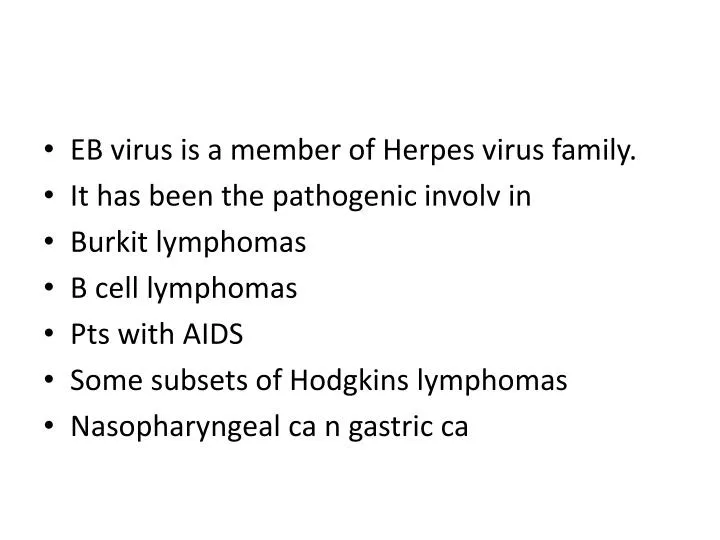
EB virus is a member of Herpes virus family. It has been the pathogenic involv in Burkit lymphomas B cell lymphomas Pts with AIDS Some subsets of Hodgkins lymphomas Nasopharyngeal ca n gastric ca. It infects the B lymphocytes n epithelial cells of oropharynx .

E N D
EB virus is a member of Herpes virus family. • It has been the pathogenic involv in • Burkit lymphomas • B cell lymphomas • Pts with AIDS • Some subsets of Hodgkins lymphomas • Nasopharyngeal ca n gastric ca
It infects the B lymphocytes n epithelial cells of oropharynx. • It induces B cell proliferation. • It uses complement receptor CD21 to attach n infect n infect the virus. • In vitro it leads polyclonalB cell proliferation and generation of B lymphoblastoid cell line
IN the endemic Burkitt lymphoma most of pts carry EB virus genome (90%) • They have elevated antibody titers against the viralantigen. • Seru antibody titers are correlated with the viral capsid antigen with the developing risk of tumor.
EB virus infects all worldwide peoples. • 15 to 20% of the people with Burkittlympoma contain EB virus apart from Africa. • There is a difference in viral genome expression with EB virus transformed B cell and Burkitt lymphomas cells. • BL doesn’t express LMP-1
In immunologically normal individuals, EBV-driven polyclonal B-cell proliferation in vivo is readily controlled, and the individual either remains asymptomatic or develops a self-limited episode of infectious mononucleosis • Burkitt lymphoma is endemic, concomitant (endemic) malaria (or other infections) impair immune competence, allowing sustained B-cell proliferation
in nonendemic areas, 80% of tumors do not harbor the EBV genome, but all tumors possess the specific t(8 ; 14) translocation • In immunosuppressed patients, including those with HIV disease and organ transplant recipients, EBV-infected B cells undergo polyclonal expansion, producing lymphoblastoid-like cells
In contrast to Burkitt lymphoma, the B lymphoblasts in immunosuppressed patients do express viral antigens, such as LMP-1, that are recognized by T cells. These potentially lethal proliferations can be subdued if the immunologic status of the host improves, as may occur with withdrawal of immunosuppressive drugs in transplant recipients
Nasopharyngeal carcinoma is endemic in southern China the EBV genome is found in all tumors. LMP-1 is expressed in epithelial cells as well. In these cells, as in B cells, LMP-1
Immune Surveillance • About 5% of individuals with congenital immunodeficiencies develop cancers, • immunosuppressed transplant recipients and patients with acquired immunodeficiency syndrome have increased numbers of malignancies • Usually Large B cell lymphomas
Most cancers occur in individuals who do not suffer from immunodeficiency. • If immune surveillance exists, how do cancers evade the immune system in immunocompetent hosts:
Several escape mechanisms are here • Selective outgrowth of antigen-negative variants. During tumor progression, • Loss or reduced expression of histocompatibility molecules • .Immunosuppression • Lack of costimulation • Antigen masking
Apoptosis by cytotoxic T cells • Eg In melenoma n Hepatocellular Ca ther is Fas expressing T lymphocytes were killed by these tumor cells n eliminate the Tumor specific T cells
Antitumor Effector Mechanisms • Cytotoxic T Lymphocytes • . In humans, they seem to play a protective role, chiefly against virus-associated neoplasms (e.g., EBV-induced Burkitt lymphoma and HPV-induced tumors). • The presence of MHC-restricted CD8+ cells that can kill autologous tumor cells within human tumors suggests that the role of T cells in immunity
Natural Killer Cells • NK cells are lymphocytes that are capable of destroying tumor cells without prior sensitization; • they may provide the first line of defense against tumor cells. After activation with IL-2, NK cells can lyse a wide range of human tumors, including many that seem to be nonimmunogenic for T cells
T cells and NK cells seem to provide complementary antitumor mechanisms • Tumors that fail to express MHC class I antigens cannot be recognized by T cells, but these tumors may trigger NK cells because the latter are inhibited by recognition of normal autologous class I molecules
Macrophages • Activated macrophages exhibit cytotoxicity against tumor cells in vitro. T cells, NK cells, and macrophages may collaborate in antitumor reactivity, because interferon-γ, a cytokine secreted by T cells and NK cells, is a potent activator of macrophages
Humoral Mechanisms administration of monoclonal antibodies against tumor cells can be therapeutically effective. A monoclonal antibody against CD20, a B cell surface antigen, is widely used for treatment of certain non-Hodgkin lymphomas.
Grading and Staging of Cancer • Methods to quantify the probable clinical aggressiveness of a given neoplasm and its apparent extent and spread in the individual patient are necessary for making accurate prognosis and for comparing end results of various treatment protocols.
For instance, the results of treating extremely small, highly differentiated thyroid adenocarcinomas that are localized to the thyroid gland are likely to be different from those obtained from treating highly anaplastic thyroid cancers that have invaded the neck organs.
Staging of cancers is based • 1. on the size of the primary lesion, • 2. its extent of spread to regional lymph nodes, • 3. and the presence or absence of metastases. • This assessment is usually based on clinical and radiographic examination.
Two methods of staging are currently in use: the TNM system (T, primary tumor; N, regional lymph node involvement; M, metastases) and • the AJC (American Joint Committee) system. In the TNM system, T1, T2, T3, and T4 describe the increasing size of the primary lesion; N0, N1, N2, and N3 indicate progressively advancing node involvement; and M0 and M1 reflect the absence or presence of distant metastases
the cancers are divided into stages 0 to IV, incorporating the size of primary lesions and the presence of nodal spread and of distant metastases. • . It is worth noting that when compared with grading, staging has proved to be of greater clinical value
Laboratory diagnosis of the cancer • Morphologic methods • Several sampling approaches are available, including excision or biopsy, • fine-needle aspiration, • and cytologic smears
Fine-needle aspiration of tumors is another approach that is widely used • . It involves aspiration of cells from a mass, followed by cytologic examination of the smear. • for breast, thyroid, lymph nodes, and salivary glands tumors are usually done with FNAC.
Cytologic (Papanicolaou) smears provide another method for the detection of cancer. • The diagnosis for the Cervical Ca (PAPsmear) • carcinoma of the cervix, often at an in situ stage
also for endometrial carcinoma, bronchogenic carcinoma, • bladder and prostate tumors, and gastric carcinomas; • for the identification of tumor cells in abdominal, pleural, joint, and cerebrospinal fluids; and, less commonly, with other forms of neoplasia.
Immunocytochemistry offers a powerful adjunct to routine histology. • Monoclonal antibodies which are specific for cellular component • Eg.Estrogen receptor for Breast Ca • Thyroglobulinfr thyroid ca • CD marker fr lymphomas • .
Flow cytometry is used routinely in the classification of leukemias and lymphomas. In this method, fluorescent antibodies against cell surface molecules and differentiation antigens are used to obtain the phenotype of malignant cells
Tumor Markers • PSA • CEA Carcinoembryonic antigen • Alpha foetoprotein CalcitoninMedullary ca of thyroid CA 125 ovarian ca HCG in choriocarcinoma
Molecular Diagnosis • Diagnosis of malignancy • Prognosis and behavior • Detection of minimal residual disease • Diagnosis of hereditary predisposition to cancer
polymerase chain reaction (PCR)-based detection of T-cell receptor or immunoglobulin genes allows distinction between monoclonal • (neoplastic) and • polyclonal (reactive) proliferations • Ewing sarcoma and several leukemias and lymphomas • BCR-ABL transcripts provides the molecular diagnosis of chronic myeloid leukemia.
Prognosis and behavior. Certain genetic alterations are associated with a poor prognosis, and thus the presence of these alterations determines the patient's subsequent therapy.
Detection of minimal residual disease. Another emerging use of molecular techniques is detection of minimal residual disease after treatment. For example, detection of BCR-ABL transcripts by PCR gives a measure of residual disease, in patients treated for chronic myeloid leukemia.
Diagnosis of hereditary predisposition to cancer. Germ-line mutation of several tumor suppressor genes, such as BRCA1, increases a patient's risk of developing certain types of cancer • detection of these mutated alleles may allow the patient and physician to devise an aggressive screening protocol, as well as to consider prophylactic surgery.
Molecular Profiling of Tumors • One of the most exciting advances in the molecular analysis of tumors has been made possible by DNA-microarray analysis. This technique allows simultaneous measurements of the expression levels of several thousand genes. The principle of this so-called gene chip technology.
Clincally important gene products and oncogenes. • 1.Tumors are Hst-1and int-2 Ca stomach,breast,bladder,melenoma • Gene product Fibroblast growth factor overexpression • 2.c myc in Burkitt lymphoma • Nuclear regulatory proteins translocations
CLINICAL ASPECTS OF NEOPLASIA • (1) location and impingement on adjacent structures, • (2) functional activity such as hormone synthesis or the development of paraneoplastic syndromes, • (3) bleeding and infections when the tumor ulcerates through adjacent surfaces,
(4) symptoms that result from rupture or infarction • (5) cachexia or wasting. • Effects of Tumor on Host • A small (1-cm) pituitary adenoma can compress and destroy the surrounding normal gland and give rise to hypopituitarism. • A 0.5-cm leiomyoma in the wall of the renal artery may lead to renal ischemia and serious hypertension..
A comparably small carcinoma within the common bile duct may induce fatal biliary tract obstruction. • The effects of Hormone production is seen with benign and malignant neoplasms arising in endocrine glands
Adenomas and carcinomas arising in the β-cells of the islets of the pancreas • Can produce hyperinsulinism, sometimes fatal. • adenomas and carcinomas of the adrenal cortex • corticosteroids that affect the patient (e.g., aldosterone, which induces sodium retention, hypertension, and hypokalemia
A tumor may ulcerate through a surface, with consequent bleeding or secondary infection. Benign or malignant neoplasms that protrude into the gut lumen may become caught in the peristaltic pull of the gut, causing intussusception and intestinal obstruction or infarction
Cancer Cachexia • suffer progressive loss of body fat and lean body mass, accompanied by profound weakness, anorexia, and anemia • cytokines produced by the tumor and the host. In patients with cancer, calorie expenditure remains high, and basal metabolic rate is increased, despite reduced food intake
TNF suppresses appetite and inhibits the action of lipoprotein lipase, inhibiting the release of free fatty acids from lipoproteins. Additionally, a protein-mobilizing factor called proteolysis-inducing factor, which causes breakdown of skeletal muscle proteins by the ubiquitin-proteosome pathway
Paraneoplastic Syndromes. • They appear in 10% to 15% of patients with cancer,1. usually earliest manifestations of occult neoplasm. • 2.Symptoms appear due to tumor or distant metastsis or hormonal changes are called as paraneoplastic syndromes. • 3.They may represent the earliest manifestation of an occult neoplasm
The most common syndromes are hypercalcemia, • Cushing syndrome, • and nonbacterial thrombotic endocarditis; • The syndromes are seen usually in theses • Lung, • breast cancers • and hematologic malignancies
The causes for. Hypercalcemia in cancer patients are as follows • the synthesis of a parathyroid hormone-related protein (PTHrP) by tumor cells. other tumor-derived factors, such as TGF-α, a polypeptide factor that activates osteoclasts, and the active form of vitamin D. • widespread osteolytic metastatic disease of bone
hypercalcemia resulting from skeletal metastases is not a paraneoplastic syndrome • Cushing syndrome • ectopic production of ACTH or ACTH-like polypeptides by cancer cells • Example is small-cell cancers of the lung
, bronchogenic carcinomas may elaborate products identical to or having the effects of ACTH, • antidiuretic hormone, • parathyroid hormone, serotonin, human chorionic gonadotropin, • and other bioactive substances
Paraneoplastic syndromes may also manifest as hypercoagulability leading to venous thrombosis and nonbacterial thrombotic endocarditis Other manifestations are clubbing of the fingers and hypertrophic osteoarthropathy in patients with lung carcinomas