Download
1 / 62
620 likes | 631 Views
Lung Cancer. Jamal Turki , M.D. The term lung cancer, or bronchogenic carcinoma, refers to malignancies that originate in the airways or pulmonary parenchyma Lung cancer is among the most common cancers worldwide

E N D
Lung Cancer Jamal Turki, M.D
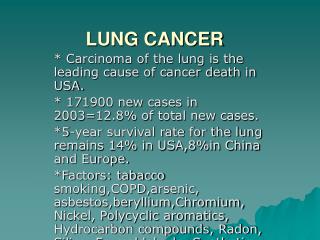
The term lung cancer, or bronchogenic carcinoma, refers to malignancies that originate in the airways or pulmonary parenchyma • Lung cancer is among the most common cancers worldwide • Lung cancer is the leading cause of cancer-related mortality in both men and women
Epidemiology • In the United States, lung cancer is the second most common cancer, after prostate cancer in men and breast cancer in women, and the most common cause of cancer deaths.
RISK FACTORS • Smoking —account for approximately 90 percent of all lung cancers • The risk of developing lung cancer for a current smoker of one pack per day for 40 years is approximately 20 times that of someone who has never smoked • prevention :In individuals who do quit smoking, the risk of developing lung cancer gradually falls for about 15 years before it levels off and remains about twice that of someone who never smoked
RISK FACTORS • Environmental toxins — These include exposure to second-hand smoke, asbestos, radon, metals (arsenic, chromium, and nickel), ionizing radiation, and polycyclic aromatic hydrocarbons • Pulmonary fibrosis — Several studies have shown that the risk for lung cancer is increased about sevenfold patients with pulmonary fibrosis • HIV infection — • Genetic factors — Genetic factors can affect both the risk for and prognosis • Dietary factors — Epidemiologic evidence has suggested that various dietary factors (antioxidants, cruciferous vegetables, phytoestrogens) may reduce the risk of lung cancer
Asbestosis & Lung Cancer(2007 American Cancer Society Data) • Prolonged heavy exposure has relative risk between 2 - 10 of causing lung cancer. • Peak incidence 15 - 24 years after exposure. • Fiber type is important: • Crocidolite & amosite > chrysotile & anthophyllite.
SCREENING Guidelines : Annual screening with low-dose computed tomography (LDCT) scanning to patients aged 55 to 74 years and who have at least a 30 pack-year smoking history and either continue to smoke or have quit within the past 15 years.
PATHOLOGY • Adenocarcinoma:40 % , Non-smokers Peripheral, Preexisting scars, Bronchoalv. • Squamous cell carcinoma- 25%, smokers, central, Hypercalcemia, cavitation • Large cell carcinoma: 5 % • Small cell carcinoma: 15 % • Others 15%
CLINICAL MANIFESTATIONS • Persons aged 50-70 years. • Lung cancer is more common in men than in women. • Symptoms may result from local effects of the tumor, from regional or distant spread, or from distant effects not related to metastases (paraneoplastic syndromes). • Approximately three-fourths of patients have one or more symptoms at the time of diagnosis.
Cough — Cough is present in 50 to 75 percent of lung cancer patients at presentation • Squamous cell and small cell carcinomas • Bronchorrhea • Post-obstructive pneumonia • bronchiectasis is uncommon
Hemoptysis : 25 to 50 percent of patients • Chest pain :same side of the chest as the primary tumor. Dull, aching, persistent pain • Dyspnea :25 percent of cases obstructive pneumonitis Atelectasis Lymphangitic tumor spread Pneumothorax Pleural effusion Pericardial effusion
Hoarseness : recurrent laryngeal nerve • Pleural involvement :typically exudates The yield of pleural fluid cytology after a single thoracentesis is about 60 percent, and the yield rises to 85 percent with three thoracenteses
Superior vena cava syndrome :sensation of fullness in the head and dyspnea. Cough, pain, and dysphagia, Physical findings include dilated neck veins, a prominent venous pattern on the chest, facial edema, and a plethoric appearance, more common in patients with SCLC than NSCLC
Pancoast's syndrome :pain (usually in the shoulder, and less commonly in the forearm, scapula, and fingers), Horner's syndrome, bony destruction, and atrophy of hand muscles.
Horner’s Syndrome • miosis (constriction of the pupils), • anhidrosis (lack of sweating), • ptosis (drooping of the eyelid) • enophthalmos (sunken eyeball)
Extrathoracic metastases • Liver • Bone • Adrenal • Brain
Paraneoplastic phenomena • Hypercalcemia:bony metastasis or secretion of a parathyroid hormone-related protein (PTHrP), calcitriol or other cytokines • SIADH secretion :SCLC, Hyponatremia • Neurologic: SCLC. Lambert-Eaton myasthenic syndrome (LEMS), cerebellar ataxia, sensory neuropathy, limbic encephalitis, encephalomyelitis, autonomic neuropathy, retinopathy, and opsomyoclonus
Hematologic manifestations — These include the following: • Anemia — Anemia is frequent in patients with lung cancer and can contribute to fatigue and dyspnea • Leukocytosis — granulocyte-colony stimulating factor • Thrombocytosis • Eosinophilia • Hypercoagulable disorders
Hypertrophic osteoarthropathy :clubbing and periosteal proliferation
Diagnosis and staging • HISTORY AND PHYSICAL EXAM • LABORATORY TESTING: complete blood count, serum electrolytes, calcium, alkaline phosphatase, albumin, (ALT), (AST), total bilirubin, and creatinine
DIAGNOSIS Chest radiographs may show the following: • Pulmonary nodule, mass, or infiltrate • Mediastinal widening • Atelectasis • Hilar enlargement • Pleural effusion
Lung Cancer:Findings on Chest X-ray • Nodule (< 3cm) vs. Mass (>= 3cm). • Location: • Peripheral (Adenocarcinoma) vs. • Central (Squamous). • Single or multiple (metastases). • Endobronchial obstruction. • Atelectasis of lobe or lung. • Pneumonia.
IMAGING • All patients with suspected NSCLC should undergo contrast-enhanced computed tomography (CT) that extends through the lungs, liver, and adrenal glands. CT is ideal for tumor node metastasis (TNM) staging • It can characterize the primary tumor and define its relationship to the chest wall and mediastinal structures • It can identify mediastinal lymph nodes that are enlarged and suspicious for malignant involvement • It can detect contralateral lung, chest wall, or upper abdominal lesions that are suspicious for metastasis
TISSUE SAMPLING • Primary tumor — There are several options for sampling a primary tumor depending on the location: • Conventional flexible bronchoscopy with forceps biopsy, blind transbronchial fine needle aspiration (TBNA), or both • CT guided TTNA • Pleural Tap • Secondary Tumor
Hypercalcemia SIADH Cushing’s Syndrome Eaton-Lambert Squamous Cell Small Cell Small Cell Small Cell Common Paraneoplastic Syndromes:SyndromeFrequent Histology
Lymph nodes • Pleural effusion • Adrenal nodule
STAGING • Based upon this initial evaluation, most patients require additional imaging. This may include whole body positron emission tomography (PET), integratedCT/PET, bone scanning, magnetic resonance imaging (MRI) of the chest wall or brain, and/or CT of the brain.
Staging for NSCLC : TNM classification Tumor size, L.Ns, Metastasis • Staging of SCLC uses the Veterans Administration Lung Study Group designations of limited (confined to one hemithorax) or extensive (beyond one hemithorax)
Treatment • Surgery • Chemotherapy • Radiation therapy
Treatment • Surgical resection offers the best opportunity for long-term survival and cure in patients with resectable NSCLC: • Lobectomy Resectability Operability
Non Small Cell Lung CancerContraindications to Surgical Resection • Stage IIIB or IV. • Extensive invasion into surrounding structures: • Vena cava or atrium involvement. • Recurrent laryngeal or phrenic nerve involvement. • SVC obstruction, malignant effusion, pericardial tamponade. • Contralateral lymph nodes.
Non Small Cell Lung CancerContraindications to Surgical Resection • Medically unfit: • Poor cardiac or pulmonary status. • Predicted postoperative FEV1% < 40%. • Predicted postoperative DLCO% < 40%. • Exercise studies for marginal candidates.