Download
1 / 1
120 likes | 492 Views
03.17 The cisterna magna septa - a potential new marker for maldevelopment of the roof of the rhombencephalon Ashley Robinson 1,2 , Ruth Goldstein 2 . 1 Children’s & Women’s Hospital of British Columbia. 2 University of California San Francisco.

E N D
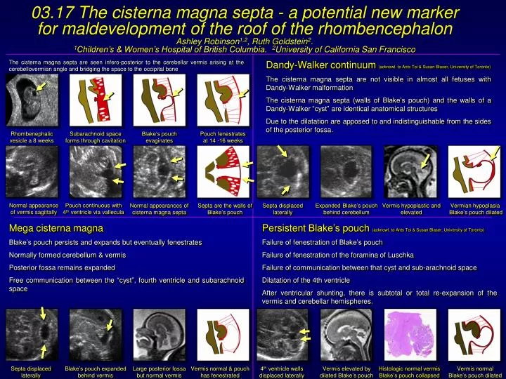
03.17 The cisterna magna septa - a potential new marker for maldevelopment of the roof of the rhombencephalon Ashley Robinson1,2, Ruth Goldstein2. 1Children’s & Women’s Hospital of British Columbia. 2University of California San Francisco The cisterna magna septa are seen infero-posterior to the cerebellar vermis arising at the cerebellovermian angle and bridging the space to the occipital bone Dandy-Walker continuum (acknowl. to Ants Toi & Susan Blaser, University of Toronto) The cisterna magna septa are not visible in almost all fetuses with Dandy-Walker malformation The cisterna magna septa (walls of Blake’s pouch) and the walls of a Dandy-Walker “cyst” are identical anatomical structures Due to the dilatation are apposed to and indistinguishable from the sides of the posterior fossa. Rhombenephalic vesicle a 8 weeks Subarachnoid space forms through cavitation Blake’s pouch evaginates Pouch fenestrates at 14 -16 weeks Normal appearance of vermis sagittally Pouch continuous with 4th ventricle via vallecula Normal appearances of cisterna magna septa Septa are the walls of Blake’s pouch Septa displaced laterally Expanded Blake’s pouch behind cerebellum Vermis hypoplastic and elevated Vermian hypoplasia Blake’s pouch dilated Mega cisterna magna Blake’s pouch persists and expands but eventually fenestrates Normally formed cerebellum & vermis Posterior fossa remains expanded Free communication between the “cyst”, fourth ventricle and subarachnoid space Persistent Blake’s pouch (acknowl. to Ants Toi & Susan Blaser, University of Toronto) Failure of fenestration of Blake’s pouch Failure of fenestration of the foramina of Luschka Failure of communication between that cyst and sub-arachnoid space Dilatation of the 4th ventricle After ventricular shunting, there is subtotal or total re-expansion of the vermis and cerebellar hemispheres. Septa displaced laterally Blake’s pouch expanded behind vermis Large posterior fossa but normal vermis Vermis normal & pouch has fenestrated 4th ventricle walls displaced laterally Vermis elevated by dilated Blake’s pouch Histologic normal vermis Blake’s pouch collapsed Vermis normalBlake’s pouchdilated