Download
1 / 68
690 likes | 1.04k Views
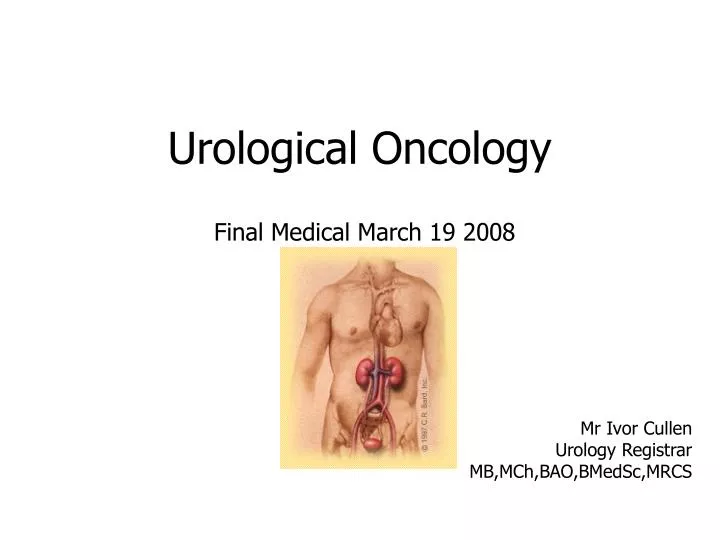
Urological Oncology. Final Medical March 19 2008. Mr Ivor Cullen Urology Registrar MB,MCh,BAO,BMedSc,MRCS. Plan. Renal Cell Carcinoma Bladder Carcinoma Testicular Carcinoma Prostate Carcinoma Miscellaneous. Renal Cell Carcinoma. 95% of Renal carcinomas AKA Clear Cell carcinoma

E N D
Urological Oncology Final Medical March 19 2008 Mr Ivor Cullen Urology Registrar MB,MCh,BAO,BMedSc,MRCS
Plan • Renal Cell Carcinoma • Bladder Carcinoma • Testicular Carcinoma • Prostate Carcinoma • Miscellaneous
Renal Cell Carcinoma • 95% of Renal carcinomas • AKA Clear Cell carcinoma • Arising from proximal tubular epithelium • Twice as common in men as in women • Occurs most commonly in the fourth to sixth decades of life • 2% assoc with inherited conditions (VHL) Cohen, Herbert T., McGovern, Francis J.Renal-Cell CarcinomaN Engl J Med 2005 353: 2477-2490
Presentation • Classic Triad – Haematuria, Flank Pain, Abdominal Mass (10%) • Most common presentations • Haematuria (40%) • Flank pain (40%) • Palpable mass in the flank or abdomen (25%) • Other signs and symptoms • Weight loss (33%) • Fever (20%) • Hypertension (20%) • Hypercalcaemia (5%) • Night sweats • Malaise • Varicocoele - usually left sided • ~ ½ cases now detected incidentally on radiographic examination
RiskFactors • Cigarette smoking doubles the risk and contributes to as many as 1/3 of all cases - dose-dependent fashion. • Obesity • Additional factors • Hypertension • Occupational exposure to petroleum products, heavy metals, solvents, or asbestos • Analgesic abuse • Acquired cystic kidney disease associated with chronic renal insufficiency • Renal dialysis • Tuberous sclerosis • Renal transplantation: With its associated immunosuppression, renal transplantation confers an 80-fold increase in the risk of renal cell cancer.
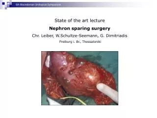
Surgical Treatment • Surgical excision is the primary treatment for organ confined RCC • Radical Nephrectomy, includes removal of the kidney en bloc with Gerota’s fascia, the ipsilateral adrenal gland,+/- regional lymph nodes is the standard therapy • Staging and evaluation for the presence of metastases, including a careful history-taking and physical examination, should be completed before surgery • Nephron-sparing Partial Nephrectomy has gained acceptance for treating tumors less than 4 cm in diameter • Renal Carcinoma Embolization • Laparoscopic Nephrectomy
Medical Treatment • Generally offered for locally advanced or metastatic renal-cell carcinoma • Immunomodulatory therapies • Interferon Alfa • Interleukin-2 • Interferon alfa and nephrectomy is superior to interferon alfa alone in metastatic disease • Chemotherapy • Rates of response to chemotherapy alone are low (roughly 4 to 6 percent)
Bladder Cancer • Median age at diagnosis is 68 years, and the incidence increases with age • Male-to-female ratio is 3:1 • 4th most common cancer in men, 10th most common in women • Almost all bladder cancers are Urothelialin origin • The urothelium consists of a 3- to 7-cell mucosal layer within the muscular bladder • Of bladder tumors, more than 90% are Transitional Cell Carcinomas TCC (Developed Countries) • ~5% are squamous cell in origin ~ 2% adenocarcinomas
Presentation • 80-90% of patients with bladder cancer present with painless gross haematuria, which is the classic presentation. • Consider all patients with painless gross haematuria to have bladder cancer until proven otherwise • Suspect bladder cancer if any patient presents with unexplained microscopic haematuria • 20-30% of patients with bladder cancer experience irritative bladder symptoms such as • Dysuria • Urgency • Frequency of urination (more advanced muscle-invasive disease or CIS) • Patients with advanced disease can present with • pelvic or bony pain, • lower-extremity oedema from iliac vessel compression • flank pain from ureteral obstruction.
Risk Factors • Smoking accounts for ~ 50% of all bladder cancers. • Nitrosamine, 2-naphthylamine, and 4-aminobiphenyl are possible carcinogenic agents • Associated with industrial exposure to aromatic amines • dyes,paints, solvents, leather dust, inks, combustion products, rubber, and textiles • Higher-risk occupations include • painting, driving trucks, and working with metal • Several medical risk factors • Patients with prior exposure to radiation treatment of the pelvis • Chemotherapy with cyclophosphamide(via acrolein) • Patients with spinal cord injuries who have long-term indwelling catheters have a 16- to 20-fold increased risk of developing SCC of the bladder • ?Weak connection between artificial sweeteners (eg, saccharin, cyclamate) and bladder cancer
Specific Work Up • Urinalysis • Urine Cytology • CT Abdo Pelvis • Upper Tract Imaging (IVP/Renal US) • Cystoscopy (Flexible/Rigid)
Treatment • Superficial Bladder Carcinoma • T.U.R.B.T ( + deep muscle biopsy) - surveillance • Intravesical immunotherapy (Bacillus Calmette-Guérin [BCG] (Ta, T1, CIS) • Induces nonspecific, cytokine-mediated immune response to foreign protein • Intravesical Chemotherapy • Epirubicin/Mitomycin C • Muscle-invasive disease (T2 and greater) • Radical Cystectomy/Cystoprostatectomy • Radiotherapy
Prognosis • Superficial bladder cancer • The risk of progression, defined as an increased tumor grade or stage, depends primarily on the tumor grade • The risk of progression increases with tumor grade, as follows: • Grade I – 10-15% • Grade II – 14-37% • Grade III – 33-64% • CIS carries a poorer prognosis and a recurrence rate of 63-92% • Diffuse CIS = ominous finding, with >70% progressing to muscle-invasive disease
Prognosis • The 5-year survival rate decreases with increasing stage, as follows: • Ta, T1, CIS – 82-100% • T2 – 63-83% • T3a – 67-71% • T3b – 17-57% • T4 – 0-22% • Prognosis for metastatic transitional cell cancer is dismal - only 5% of patients living 2 years after diagnosis • Early diagnosis and improvements in treatment of bladder cancer may be responsible for the improved survival rate of patients with TCC
Testicular Cancer • Overall testicular cancer is not very common (2000 new cases UK/year – 1% all cancers) • Primarily affects young men - 20 to 44 where it is the most common cancer • Testicular cancer is curable in the majority (over 90%) of cases
Presentation • Painless unilateral swelling • Scrotal swelling after minor trauma • Scrotal / Lower abdo pain • Hydrocoele • Endocrinological Effects – • Gynaecomastia / Breast tenderness • Decreased libido • In 10% presenting symptoms due to metastatic disease • Neck mass • Cough/Dyspnoea • GI / back / bone pain
Classification • Germ Cell Tumours (95% of all) • Seminomas (40% of germ cell tumours) • Non Seminomatous (60% of germ cell tumours) • Most nonseminomas contain cells from at least two subtypes, including the following: • Choriocarcinoma (rare; aggressive; likely to metastasize) • Embryonal carcinoma (accounts for 20% of cases; likely to metastasize) • Teratoma (usually benign in children; rarely metastasize) • Yolk sac carcinoma (most common in young boys; rare in men) • Non Germ Cell Tumours (5% of all) • Leydig Cell Tumours • Sertoli Cell Tumours • Others
Risk Factors • Age • Cryptorchidism • 3-5% chance of cryptorchid testis developing cancer • Family History • Race • ? Trauma • ? Orchitis
Work Up • Serum tumor markers • At the initial presentation of a patient with a testis tumor • Serum human chorionic gonadotrophin (βHCG), alpha-fetoprotein (AFP), and lactate dehydrogenase (LDH) are the most important tumor markers. • Following markers to assess success of treatment • AFP has a half-life of 5-7 days, and HCG has a half-life of 36 hours • Ultrasound • Most tumors are diagnosed based on physical examination finding • Performed to ensure the correct diagnosis or to establish a diagnosis in a patient in whom the testicular examination cannot differentiate the scrotal structures • In the setting of teratoma elements, ultrasound images may demonstrate well-defined structures of ectodermal derivation
Work Up • CT scan • CT scan of the abdomen and pelvis is integral to the staging of a testis tumor • Left-sided NSGCTs typically spread first to the left para-aortic and then preaortic lymph nodes inferior to the renal vessels • Right-sided tumors spread to the paracaval and interaortocaval nodes inferior to the renal vessels. • A chest radiograph or CT Thorax is usually obtained to help identify any possible pulmonary metastases • Each patient should be offered the opportunity to obtain a semen analysis and to bank his sperm for future fertility concerns • This can be performed either before or after the orchiectomy • The treatment options can significantly impact future fertility
Staging TNM classification • pT0 no evidence of primary tumour, e.g. histological scar in testis • pT is carcinoma in situ (CIS, TIN) • pT1 tumour limited to the testis and epididymis without vascular/lymphatic invasion; tumour may invade into the tunica albuginea but not the tunica vaginalis • pT2 tumour limited to the testis and epididymis with vascular/lymphatic invasion or tumour extending through the tunica albuginea with involvement of the tunica vaginalis • pT3 tumour invades the spermatic cord with or without vascular/lymphatic invasion • pT4 tumour invades the scrotum with or without vascular/lymphatic invasion.
Treatment • Complicated….. • Depends on TNM • In general – • Localized Seminoma • Inguinal Orchiectomy +/- Radiotherapy to Nodes • Seminoma with nodes • Inguinal Orchidectomy + Platinum based chemotx • Nonseminomatous tumour • Inguinal Orchidectomy +/- RPLND +/- Chemotx
Introduction • 1400 cases of adenocarcinoma of prostate in Ireland per year – (Most commen solid tumour in men) • A large proportion die from it – 580 deaths per year in Ireland • Also has a significant morbidity rate • Palliation only treatment available for advanced disease –(2/3 men metastatic at presentation) • Detection and treatment of organ-confined disease remains the only hope for cure
However • Limited sensitivity to current screening tools • Influence of screening on survival subject to bias • Scarcity of reliable markers for predicting progression • Considerable morbidity associated with curative treatments
Incidence of Prostate Cancer • Histological – 30% of men age 50 • Spread • Direct – Bladder / Seminal Vesicles • Lymph nodes – Pelvic and Paraaortic • Blood – Prostatic venous plexus to vertebral venous plexus • Clinical – Lifetime risk 16.7% • Death from prostate cancer - Lifetime risk 2.5%
Signs & Symptoms • Often asymptomatic • Symptoms of lower urinary tract obstruction may not be present • Hesitancy, poor stream, nocturia. • Post renal failure, uraemia & confusion • Bony pain • Anaemia • Haematuria (Due to BPH in 90% cases) • DRE – May have firm, nodular prostate
Risk Factors • Age • Family History • Race • Diet high saturated fat • ? Vitamin Deficiencies (D,E)
Initial tests • FBC – ?anaemia • U&E – check renal function • LFT – Alk phos • MSU - ?haematuria / concurrent UTI • PSA…
PSA 1 • Cheap • Widely available • Acceptable to most men • Easy to interpret However • Not prostate cancer specific • Normal value does not rule out cancer • Many patients fall into ‘grey area’
PSA 2 • Before taking test, ensure… • 1 No urological instrumentation x 1/52, including catheters, but not DRE. • 2 No ejaculation x 48 hrs • 3 No Bicycle riding x 1/52 • 4 No current UTI
PSA 3 • Many attempts to increase PSA sensivity • Age adjusted PSA • PSA density • PSA velocity / PSA doubling time
Recommendations American Cancer Society Do not recommend mass screening, but men should be given the opportunity for shared decision making about testing Annual PSA & DRE from 50 years (45 in higher risk groups) American Medical Association Mass screening premature Annual PSA & DRE from 50 years (40 in higher risk) United States Preventive Services Task Force Insufficient evidence to recommend for or against screening National Health Service Screening will not be offered until there is clear evidence that screening will bring about more benefit than harm Ireland Screening recommended (RCSI guidelines)
Radiological Investigations • Pelvic / Lumbar spine xray • ?osteosclerotic lesions • Renal USS (if raised renal profile) • TRUS biopsy • Invasive • Not definitive, may need to be repeated • Debate over number of core biopsies • Bone scan • In presence of PSA>20 / bony pain • MRI prostate – poor specificity without endorectal coil
Staging • pT1 – asymptomatic, no clinical signs • pT1a <5% TURP chippings • pT1b >5% TURP chippings • pT1c raised PSA indicating TRUS biopsy • pT2 – Palpable, confined to prostate • pT2a <2cm, one lobe • pT2b >2cm, one lobe • pT2c Any size, both lobes • pT3 – Locally Invasive • pT4 – Distant metastasis
Gleason Grade • Histological grading of prostate cancer 1-5 However • Prostate cancer not uniform • To aid calculations of prognosis, the sum of the 2 most prevalent islands of prostate cancer are used • Therfore, gleason grade ranges 2-10
Treatment Options • Stage 1 or 2 disease • Radical Prostatectomy • Radiotherapy • Stage 3 or 4 disease • Hormonal Therapy • Watchful Waiting
Radical Retropubic Prostatectomy • Prerequisites • Confirmed histological diagnosis • At least 10yrs life expectancy post procedure • PSA < 20ng/ml • Gleason grade <8 • Negative bone scan +/- negative MRI • Patient fully counselled and aware of possible complications and alternative treatment options