Download
1 / 35
350 likes | 479 Views
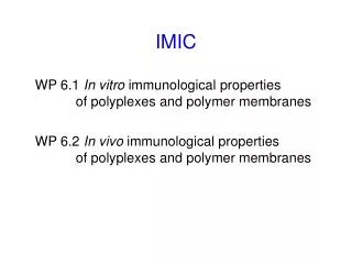
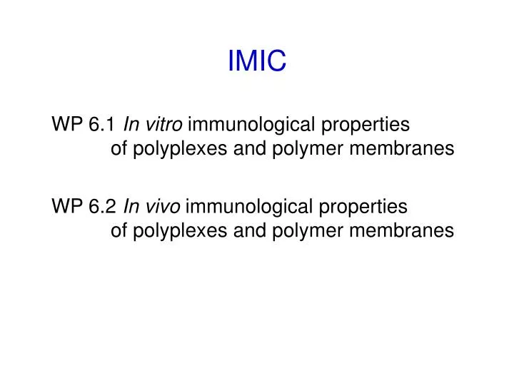
IMIC WP 6.1 In vitro immunological properties of polyplexes and polymer membranes WP 6.2 In vivo immunological properties of polyplexes and polymer membranes. SUMMARY: Preincubation of hydrogels is important: optimal protocol was found

E N D
IMICWP 6.1In vitro immunologicalproperties of polyplexes and polymer membranesWP 6.2In vivoimmunological properties of polyplexes and polymermembranes
SUMMARY: Preincubation of hydrogels is important: optimal protocol was found Gelatin B does not induce spontaneous hemolysis of human ery Spontaneous proliferation of mouse spleen cells and human PBMC is not increased within 3 days of cultivation on hydrogel Gelatin B hydrogel does not induce NO production by mouse macrophages - no inflammatory stimulus Gelatin B does not induce any significant toxic damage to cells of different origin (normal and permanent cell lines) Growth and viability of cells (normal, permanent cell lines) is modulated due to hydrogel surface (clusters)
Biocompatibility and immunocompatibility of the gelatin B hydrogel To finish the tests with gelatin hydrogels and to conclude the results: Cytokine detection in supernatants FLOW CYTOMIX: IFNgamma, IL-1alpha, IL-2, IL-4, IL-5, IL-6, IL-8, IL-10, IL-12p70, TNFalpha, TNFbetaβ samples: supernatants of mouse spleen cells, unstimulated and mitogen-stimulated; PEC mRNA quantification (Riboquant) for selected cytokines Other types of hydrogels could be studied using the same protocols
Cationic polymers for preparation of polyplexes, plasmids • in vitro assays (preliminary results) • hemolysis (control of biocompatibility) • proliferation of normal cells • polyplexes - in vitro: proliferation of normal cells, permanent cell lines • in vivo study: immunization (i.p, s.c.) • PEC - cellularity, phenotype, cytokines, heat shock proteins • antibody production (anti-DNA, anti-polymer, anti-polyplex) • genotype – Th1/Th2 prone (HLA-DR4, DR5)
In vitro screening - hydrogels • Hemolysis • Proliferation of mouse spleen cells • Proliferation of human peripheral blood mononuclear cells • Cultivation of mouse peritoneal cells • Growth of permanent cell lines • endothelial • fibroblast • monocyte/macrophage • Cytokine production by cells cultivated on hydrogels
Gelatin B-hydrogels: mitogen-stimulated mouse spleen cellsunstimulated = ctrl Supernatants: 24 h, 48 h for cytokine detection T cell stimulation: ConA T and B stimulation: PWM B cells, monocytes/macrophages: LPS FlowCytomix assay (BenderMedsystems) IL-1α, IL-2, IL-4, IL-5, IL-6, IL-10, IL-17, GM-CSF, IFNγ, TNFα Th1 type responses: high IFNγ, IL-12, TNFα, low IL-10 Th2 type responses: IL-4, IL-5, high IL-10 mRNA samples stored (Riboquant)
FlowCytomix bead-based assays follow the same principle as a sandwich immunoassay (ELISA) quantitative assay Fluorescent polystyrol beads are coupled with antibodies specific to the analytes to be detected. A mix of coupled beads is incubated with the samples to be tested. Analytes in the sample bind to the antibodies coupled to the beads. A biotin conjugated antibody mix is added, which binds to the analytes bound to the capture antibodies. Streptavidin-Phycoerythrin (PE) is added which binds to the biotin conjugates. Beads are differentiated by their sizes and distinct spectral signature by flow cytometry. FlowCytomix assay enables calculation of analyte concentration in the tested samples.
FlowCytomix assay: quantitative multiplex for 10 cytokines Biotin-conjugated antibody Vead coupled with antibody Streptavidin-PE
25 ul sample IL-1α: 15.7 pg/ml IL-2: 8.8 pg/ml IL-4: 0.7 pg/ml IL-5: 4.0 pg/ml IL-6: 2.2 pg/ml GM-CSF: 10.9 pg/ml IFNγ: 0.8 pg/ml IL-10: 13.3 pg/ml IL-17: 2.4 pg/ml TNFα : 0.9 pg/ml
SUMMARY: Prolonged preincubation of hydrogels is necessary Gelatin B does not induce spontaneous hemolysis of human ery Spontaneous proliferation of mouse spleen cells and human PBMC is not increased within 3 days of cultivation on hydrogel Mitogen-induced proliferation of mouse spleen cells and human PBMC is rather decreased, but SI remain comparable Gelatin B hydrogel does not induce NO production by mouse PEC and mouse macrophage cells RAW, and does not induce TNF production by human THP-1 cells Growth of permanent cell lines is rather limited in monocyte /macrophage cell lines (RAW, THP-1), and almost unchanged in fibroblasts (3T3) and endothelial cells (EAHY) Density of cell seeded on the hydrogel is important Growth and viability of cells (normal, permanent cell lines) is modulated due to the hydrogel surface (clusters)
SUMMARY: Prolonged preincubation of hydrogels is necessary Gelatin B does not induce spontaneous hemolysis of human ery Spontaneous proliferation of mouse spleen cells and human PBMC is not increased within 3 days of cultivation on hydrogel Mitogen-induced proliferation of mouse spleen cells and human PBMC is rather decreased, but SI remain comparable Gelatin B hydrogel does not induce NO production by mouse PEC and mouse macrophage cells RAW, and does not induce TNF production by human THP-1 cells Growth of permanent cell lines is rather limited in monocyte /macrophage cell lines (RAW, THP-1), and almost unchanged in fibroblasts (3T3) and endothelial cells (EAHY) Density of cell seeded on the hydrogel is important Growth and viability of cells (normal, permanent cell lines) is modulated due to the hydrogel surface (clusters)
Sample Description:Methacrylamide modified gelatin (type A) Structure / Product Information: Gel-MOD type A, 1 equivalent methacrylic anhydride (DS 70%), uncrosslinked, freeze-dried form, sterilized using ethylene oxide Sample Description: Methacrylamide modified gelatin (type B) Structure / Product Information: Gel-MOD type B, 1 equivalent methacrylic anhydride (DS 67%), uncrosslinked, freeze-dried form, sterilized using ethylene oxide Sample Description: Irgacure 2959® Structure / Product Information: 1-[4-(2-Hydroxyethoxy)-phenyl]-2-hydroxy-2-methyl-1-propane-1-one (Ciba Speciality Chemicals N.V.), photo-initiator, sterilized using ethylene oxide
Endothelial cells: EAHY Viability: Alamar blue, 48 h Cellular damage: lactate dehydrogenase (LDH)
Human mononuclear cells THP-1 Viability: Alamar blue, 48 h Cellular damage: lactate dehydrogenase (LDH)
Mouse macrophages RAW 264.7 Viability: Alamar blue, 48 h Cellular damage: lactate dehydrogenase (LDH)
Mouse macrophages RAW 264.7 nitric oxide (NO) production (24 h) nitrite (uM)
Mouse macrophages RAW 264.7 Viability: Alamar blue, 24 h
? Endotoxin contamination Limulus amoebocyte lysate assay ?? polymerized gelatin A to compare with the gelatin B
Gelatin A hydrogel mechanical properties of type A gelatin are worse – soft, floating … Hemolysis: no spontaneous hemolysis
Gelatin coated glass slides Sample Description:Gelatin (type B, gel-MOD) coated glass slides in 24 well plates Structure / Product Information: Half of the wells have been filled with glass slides (Ø 15 mm) spincoated with gelatin type B. The other half is left empty for the controls. The gelatin films have been prepared by polymerization of methacrylamide modified gelatin using a UV-active photoinitiator (i.e. Irgacure 2959®) and applying UV-light (276 nm, 10 mW/cm2) to the spincoated glass slides for 20 minutes.
Laser scanning microscopy RAW 264.7 cells: number of living cells on the gelatin coated glass and on the plastic surface was comparable (24 h); almost no dead cells !! Cells grow on both sides of the glass
Sample Description: Gelatin coated polyimide membranes Structure / Product Information: polyimide + aminopropylmethacrylamide + 5 w/v% gel-MOD, incubated in double distilled water at 37°C during two hours, after UV-irradiation during 30 minutes Sample Description: Polyimide membranes modified with AFB Structure / Product Information: polyimide + AFB (5 mg/ml) in ethyl acetate overnight, followed by UV-C irradiation (250 nm) for 20 minutes
Cationic polymers for preparation of polyplexes, plasmids • in vitro assays (preliminary results) • hemolysis (control of biocompatibility) • proliferation of normal cells • polyplexes - in vitro: proliferation of normal cells, permanent cell lines • in vivo study: immunization (i.p, s.c.) • PEC - cellularity, phenotype, cytokines, heat shock proteins • antibody production (anti-DNA, anti-polymer, anti-polyplex) • genotype – Th1/Th2 prone (HLA-DR4, DR5)
Kationic polymers and polyplexes • Hemolysis and hemagglutination • In vitro: selected cell lines (EAHY, THP-1, RAW) and normal cells (mouse spleen cells) = incubation – viabiliy and cellular damage (LDH)Supernatants: cytokine detection by FlowCytomix • In vivo: injection of polyplexes i.v. – serum samples for cytokine detection after 8, 24 and 48 (72 hours) • To compare the polymers: calf thymus DNA? • Selected polymers with the plasmid