Download
1 / 40
400 likes | 624 Views
Immunology 4 Specific immunity - molecules . Immunoglobulines Complement MHC Cytokines. Molecules of specific immunity. some are common for nonspecific immunity, too

E N D
Immunology 4Specific immunity - molecules • Immunoglobulines • Complement • MHC • Cytokines
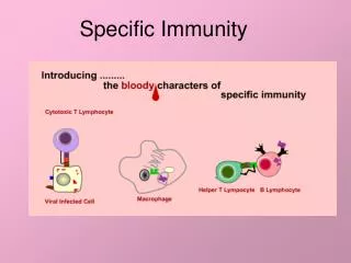
Molecules of specific immunity • some are common for nonspecific immunity, too • others only for specific immunity (BCR, TCR = antigen specific receptors on B resp. T cells) • T cells receptor - TCR, B cell receptor - BCR • TCR bind specific antigen, bound on molecule MHC • BCR bind specific antigen directly • B cells synthetise immunoglobulins • synthesis of cytokines after activation of T and B cells
Differences • Nonspecific immunity - system prepared before exposition antigen - not differe quality and type of antigen - can be strenghten via cytokines after exposition to antigen • Specific immunity - system prepared before exposition, but induced by exposition to antigen - lag phase - strenghten by antigen and in next exposition - slight differentiation of antigens • Major signs of specific immunity are memory and specificity
ANTIBODIES Blood serum of immunised annimals contains specific molecules – antibodies, that bind antigen, that induced their production Behring, Kitaso 19th century Serum– liquid part after centrifugation of clotted blood Plasma– liquid part after short centrifugation of not clotted blood Serum containing antibodies against specific antigen – antiserum Antibody – molecule of immunoglobuline with specificity for the epitope.
FUNCTION OF ANTIBODIES • specific binding of antigen and neutralisation of its function - this binding results in other reactions that lead to - elimination of antigen (activation of complement, macrophages, opsonisation in phagocytosis). • Reaction of antigen and with antibody differs according to the type of antigen *corpuscular antigens – microbes, erytrocytes – agglutination *solubil antigens – immunocomplexes in solution - precipitation
Immunoglobulins • Synthetised by B lymphocytes (B cells) • present in cytoplasma and on membrane B bb • synthetised and secreted by plasmatic cells Structure Isotypes
Structure of immunoglobulins • 4 polypeptides – 2 identical light chains (LC) - 2 identical heavy chains (HC) bound by disulfidic binding • NH end of one light chain and one heavy chain form the epitop binding site
Structure of immunoglobulins HC and LC are divided to identical domains • LC contains 2 domains - 1 constant CL - 1 variable VL HC contains 4 or 5 domains - 1 variable VH - 4 or 5 CH epitope binding site
Štructure of immunoglobulines • Light chain – kappa (k) or lambda (l) - one palsmatic cell produce only one type of light chain • Haevy chains – isotypes • – mi (m) - IgM • - epsilon (e)- IgE • - gama (g) - IgG • - alfa (a) - IgA • - delta (d) - IgD
IgG Existuje ako povrchový aj secernovaný monomér 2 g + 2k (alebo 2l) Existujú 4 podtypy ťažkých reťazcov a teda 4 IgG podtriedy: IgG1 IgG 2 IgG3 IgG4 - Najviac v sére, - aktivácia komplementu, - opsonizácia, - neutralizácia mikroorganizmov, - ADCC, - hypersenzitívne reakcie (II., III.,), - prechod transplacentárnou bariérou, - protektívne
IgM • bound as monomer on cell surface • or in serum and liquids as pentamer with 10 H a 10 L chains bound in paire by disulfidic bindings and J chains. • First synthetised after stimulation (in utero, too) • Immobilisation of antigen • activation of C´ by classical path
IgA • monomer in seurm • dimer (2 monomers + J chain) • mucuse – translocation through membrane bind on receptor that bind on dimer as SC (secretion component) – that gives resistence to ensymatic degradation - saliva, tears, mucous, breast milk, GIT secretion • IgA1 - serum and secretion up diaphragma IgA2 – secretion epitelium, GIT, respiratory trakt • Mostly produced
IgD • Monomer • Almost the only role is the role of receptor for antigen on the surface of B cell • Other functions are not known • Contain the intercelular part - tail piece
IgE • in low concentration in serum • mostly on the surfaces of mastocytes, monocytes and eosinofils and basofils • Specific receptors for Fc fragment of IgE molecule (FCeRI). Mastocytes are bound by antigens on Fab phragments that leads to degranulation and the release of histamin lead to alergic reactions.
B cell • produces immunoglobulines with light chain kappa or lambda • produces only one isotype (heavy chain) • nonstimulated B cell has on the surface the molecule of monomer of IgM or IgD as BCR • After secretion to tissues the IgG, IgE will be monomer., IgM – as soluble pentamer, IgA - soluble dimer and monomer
Complement • Consists of innactive circulating glycoproteins activated in cascade way after innitial stimulation • 3 paths of stimulation: - classic ( antigen + antibody) - alternative ( microbes and their products) - MBL path • Results in production of: MAC membrane attact complex it is bound on cell surface (of microbe) formation of disruptions in surface memgrane that leads to lysisof cell
Activation of classical pathway C´ • Ag + Ab = production of MAC, opsonisation (C3b), production of anaphylatoxins (C3a, C5a, C4a) • Activation C1: - IgM (IgG) + Ag = konformationnal change of Fc = binding of C1q on CH2 (Fc fragment of Ig molecule). - Subsequently binding of C1r and C1s - C1qrs has ensymatic activity and disrupt C4, C2, C3 on fragments C4b2b3b, that is C5covertase – beggining of MAC
MHC – major histocompatibility complex • Named also HLA complex = segment on chromosome 6 containning several genes needed for good function of immunity (6.7). These genes encode ensymes and structural molecules needed for activation and function of T and B cells. These are molecules of classes I, II a III MHC (resp.HLA).
MHC • Surface molecules • MHC class I, II and III (C4,Bf C2 – part of complement) • MHC I - 3 domaines – polypeptide chains and beta2 microglobulin • presentation of endogenous antigens to T lymphocytes. • present on all cells with nucleus • MHC II - alfa a beta polypeptid chains • presentation of exogennous antigens - present on macrophages, B lymphocytes and some other antigen presenting cells - APC)
MHC I – on cells with nucleus • polymorphisme HLA – A, - B, - C with more than 100 allels for each locus • in heterosygots: one of 6 different types of molecules of MHC I (AA, AB, BB, BC,CC, AC) can be exprimed on individual cell • MHC I forme cleft between a1- a2 domaines, that bind 8-9 aminoacides peptid. • There are minor MHC Ib (HLA E, -F, -G, -H locuses)
MHC II – on APC cells • dendritic, macrophage, B cells, some activated T cells, specialised epitelial cells in thymus and intestine. • Heterodimer of a1 chain and b1chain • Encoded in HLA-DP, -DQ, -DR segment of 6th chromosomes • After synthesis a combines only with b chain of the same locus (DPa to DPb) on the same (cis) or other (trans) member of chrmosome pair • Enable a big variability of dimers of MHC II in heterosygots
T cell receptor, TCR Antigen specific receptor on T cell Heterodimer peptid pair: ab, gd Differenciation of the type of heterodimer peptid pair is done early during development of T cell Contain variable (Va, Vb., orVg, Vd) and constant Ca, Cb., orCg, Cd) part of domains.(like in Ig molecule) Each T cell has unique TCR (like in Ig on B cells) TCR recognise antigen only in cleft of MHC (differ from Ig, that recognise soluble peptides)
T cell receptor - TCR Responsible for diversity of antigen specificity (recognises) 1015 epitopes • Interaction of antigen connected to MHC II on APC – antigen presenting cell – and TCR leads to transmission on signal via CD3 and activation of T cells for next reactions.
Molecules of cell interaction Interaction is performed: • by direct intercellular contacts • signals (soluble molecules) secerned, sent and received by others Immunocompetent cells positively or negatively regulate their functions, migracte to specific places, make vital decisions. Cytokines, Chemokines, Adhezsines, CD molecules, Signla transmission molecules
Cytokines Biologically active substances released from specific cells influencing other cells (intercellular mediators, signals) lymphocytes - lymphokines, monocytes - monokines, interleukines – communiction between leu autocrine – influence the same cells, paracrine – influence narrow, by site cells Secreting is short-time and limited Influence releasing and production of other cytokines – waterfall (cascade) activation and stimulation Agonistic or antagonistic action
Cytokines acc. function Mediators and regulators of nonspecific immunity - TNF - tumor necrosis factor, interleukín -IL 1, IL 10, interferon gama IFN-gama Mediators and regulators of specific immunity -IL 2, IL 4, IL 5, IL 10, IFN gama Stimulators of hematopoesis - IL 3, colony stimulating factors CFS
Other cytokines, molecules • Chemokines – chemoattraction based on chemical gradiet, chemotaxis • Adhesive molecules – produce contact between cells (direct intercellular) – integrines, selectines, addressines– tissue tropismus • CD – Cluster of differentiation molecules – on surfaces of different cells – indicators of functional capacity of cells CD3 – transmission of signal throught cell membrane after activation of TCR CD4 – on 2/3 T cells, Thelper, recognise antigen in MHC II CD8 - on 1/3 mature T cells, T supressoric, T cytotoxic, recognise antigen in MHC I
Signal transmitting molecules • Leu use surface receptors to patrol extracellulare space • Enganement of receptor and ligand communicate signal in the cell throught the cytoplasmatic tail, that starts the cascade of chemical signals, that regulate transkription of genes in nucleus that is responsible for changes of cell activity. • (example: molecules of JAK-STAT, Ras-MAP path)