Download
1 / 83
840 likes | 1.46k Views
Important developments of the Vertebrates: brain and sense organs. The ancestors of vertebrates switched from filter feeding to more active feeding, which required movement and the ability to sense the environment in detail. Important developments of the Vertebrates: brain and sense organs.

E N D
Important developments of the Vertebrates: brain and sense organs • The ancestors of vertebrates switched from filter feeding to more active feeding, which required movement and the ability to sense the environment in detail.
Important developments of the Vertebrates: brain and sense organs • The need to gather and analyze information led to the development of multiple sense organs among the vertebrates. • These include complex eyes, pressure receptors, taste and smell receptors, lateral line receptors for detecting water vibrations, and electroreceptors that detect electrical currents.
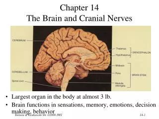
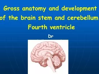
Important developments of the Vertebrates: brain and sense organs • The development of sensory structures and increased mobility generated the need for a control center to process information. • The anterior end of the nerve cord consequently became enlarged into a brain.
Important developments of the Vertebrates: brain and sense organs • The vertebrate brain in fact developed into a tripartite brain (with a forebrain, midbrain, and hindbrain) that was enclosed within a protective cranium of bone or cartilage.
Brain structure • In the most primitive forms of brains the forebrain is associated with the sense of smell, the midbrain with vision and the hindbrain with balance and hearing. • From this primitive condition the size and complexity of the brain has greatly increased.
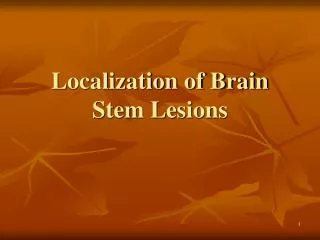
Hindbrain • The Hindbrain has two portions • Most posterior portion is the Medulla oblongata. It operates primarily at the reflex level. Reflex centers for respiration, heartbeat, and intestinal movement are found in the medulla oblongata. • The medulla oblongata also relays signals from the inner ear and is a major pathway through which signals pass to and from higher areas of the brain. • Damage to the medulla, not surprisingly, is life-threatening.
Hindbrain: cerebellum • The anterior portion of the hindbrain includes the cerebellum (present only in jawed vertebrates), which is highly folded and convoluted. • The cerebellum integrates sensory information (touch, vision, positional, hearing) with motor input to maintain the organism’s equilibrium (its position and equilibrium in relation to gravity). • The cerebellum also coordinates motor movements, both reflex movements and directed movements.
Hindbrain: cerebellum • If the cerebellum is removed an organism’s movements become uncoordinated and uneven. • In humans damage to the cerebellum causes a condition called dysmetria in which someone reaching for a target with their hands (or feet) overshoots or undershoots it.
Cerebellum • The size of the cerebellum is proportional to its role. • In fish, the cerebellum is proportionally enlarged in part because it must process lots of input from the lateral line system, but also because fish must orient themselves in three dimensions and equilibrium and balance are thus very important. • In bottom-dwelling fish and those that are not active swimmers the cerebellum is relatively small.
Midbrain • The midbrain develops in step with the eyes and is the part of the brain that receives visual information. • The roof of the midbrain (the tectum) is the part that receives visual information (and also lateral line and auditory input). The floor of the midbrain (the tegmentum) initiates motor output based on the input received. • The midbrain is often the most prominent portion of the brain in fish and amphibians
academic.emporia.edu/sievertl/verstruc/fbrain.htm Frog brain model
Forebrain: diencephalon and telencephalon • The forebrain has two major parts the posterior diencephalon and the anterior telencephalon. • The diencephalon includes the pineal gland, pituitary gland, thalamus and the hypothalamus.
Diencephalon: Hypothalamus • The hypothalamus plays a major role in homeostasis, the regulation of the body’s internal physiological balance including such aspects as temperature, water balance, appetite, blood pressure and sexual behavior. • It achieves this by the release of hormones either produced in the hypothalamus itself or stimulating the release of hormones from the pituitary gland.
Diencephalon: Pituitary Gland The posterior pituitary gland stores and releases two hormones produced by the hypothalamus: oxytocin and antidiuretic hormone (ADH). In humans oxytocin stimulates uterine contractions in childbirth and also milk production. ADH acts on the kidneys to increase water retention and reduce urine volume.
Diencephalon: Pituitary Gland • The anterior pituitary (AP) produces many hormones that in turn control the activity and hormone release from other endocrine glands including the gonads, adrenal glands, and thyroid. • Growth hormone is produced in the AP and has a direct effect on growth. Excess production can lead to gigantism and deficient production to dwarfism.
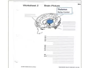
Diencephalon: pineal gland and thalamus • The pineal gland affects skin pigmentation by affecting melanocytes. It also plays a role in regulating biological rhythms. • The thalamus is the major coordinating center for incoming sensory impulses from all over the body and it relays the information to the cerebral cortex.
Telencephalon • The telencephalon (or cerebrum) includes two expanded lobes, the cerebral hemispheres (which in many mammals are greatly folded) and the olfactory bulbs. • The receipt of olfactory information is a major role of the telencephalon and in species in which olfactory information is important the olfactory bulbs are greatly enlarged.
Cerebral hemispheres • The cerebral hemispheres in reptiles and especially in birds and mammals are enlarged 5 to 20-fold over those of non-amniotes of comparable size. • The enlarged size of the cerebral hemispheres allows more and faster processing of sensory information and thus greater intelligence.
Cranial nerves • Not all nervous inputs and outputs to and from the brain travel via the spinal cord. • A series of cranial nerves connect directly into the brain. • Most have Roman numerals for names and many of them directly connect to sensory structures and other structures in the head.
Cranial nerves • The cranial nerves are: • Cranial nerve 0: Nervus terminalis runs to blood vessels of the olfactory epithelium. • Cranial nerve I: Olfactory nerve: connects with olfactory cells in the mucous membranes of the olfactory sac. • Cranial nerve II: Optic nerve: connects to the eyes. • Cranial nerves III and IV and VI: connect to extrinsic eye muscles. • Cranial nerve V: Trigeminal nerve: branches into three nerves that connect to the eye, jaws and the skin of the head. Cranial nerve VII also innervates the face as well as the taste buds.
Cranial nerves • Cranial nerve VIII Auditory nerve connects to the inner ear. • Cranial nerve IX: Glossopharyngeal nerve connects to taste buds and parts of the throat. • Cranial nerve X: Vagus nerve: serves areas of the mouth, pharynx and most of the viscera. • Cranial nerve XI: supplies some jaw muscles and the trapezius. • Cranial nerve XII: Hypoglossal nerve innervates tongue muscles • Cranial nerves arise from both neural crest cells and from ectodermal placodes in the embryo
Neural crest cells • Neural crest cells are groups of special cells derived from the embryonic dorsal tubular nerve cord. • Early in development these cells separate from the neural tube before it closes. • They assemble into cords above the neural tube and migrate along distinct pathways to various permanent locations where they differentiate into a variety of structures.
Image Source: http://www.niaaa.nih.gov/publications/arh25-3/175-184.htm
Neural crest cells • Neural crest cells give rise to among other structures: • Schwann cells • Some components of the peripheral nervous system • Odontoblasts (give rise to dentin) • Dermis of facial region (from which many skull bones are produced) • Beak of birds • Some chromatophore cells • Connective tissue of the heart • Parts of the meninges
Ectodermal Placodes • Ectodermal placodes (with some exceptions in fish) are thickenings of the surface ectoderm that sink inwards and develop into various sensory structures. • Paired olfactory placodes that form at the tip of the head develop into odor receptors that connect to the brain. • Paired optic placodes produce the lens of the eye.
Ectodermal Placodes: Vestibular apparatus • Some dorsolateral placodes (in fish) give rise to the lateral line system. • The otic placode (one of the dorsolateral placodes) forms the vestibular apparatus in the inner ear. • The vestibular apparatus plays a major role in both balance and hearing.
Ectodermal Placodes: Vestibular apparatus • There are three semicircular canals (arranged at roughly 90 degree angles to each other) and two connecting structures (the sacculus and utriculus) in the vestibular apparatus • The canals are fluid filled and respond to rotation when the head is tilted. The information about orientation and motion is then delivered to the brain for interpretation.
http://goodrich.med.harvard.edu/pictures/BRODEL34smaller.bmp
Ectodermal Placodes: Vestibular apparatus • In some fishes and in reptiles, birds and mammals a section of the vestibular apparatus (the lagena) is specialized for sound reception. • In terrestrial vertebrates the lagena is usually elongated and in most mammals it becomes coiled forming the cochlea.
Ectodermal Placodes: Vestibular apparatus • In mammals sound vibrations are transferred from the eardrum via the inner ear bones (malleus, incus and stapes) to the cochlea. • The vibrations cause hair cells in the fluid filled cochlea to move and this movement is converted into nerve signals that are then transmitted to the brain where they are interpreted as sounds.
Significance of neural crest cells and ectodermal placodes • The vertebrate head is mostly a collection of parts that are derived from neural crest or ectodermal placode tissue. • These unique tissues and their mode of embryonic production distinguish vertebrates from all other chordates.
The role of hox genes in the evolution of the Vertebrates • A factor that may have played a role in the evolution of the vertebrates is the duplication of the Hox gene complex. • Hox (short for hemeobox) genes are master control genes that regulate the expression of a hierarchy of other genes during development.
Hox genes • Because a single hox gene influences the expression of many other structural genes a change in when and where a hox gene is turned on may lead to major morphological changes in the phenotype such as the addition or loss of legs, arms, antennae and other structures.
http://evolution.berkeley.edu/evolibrary/images/mutantfly.jpghttp://evolution.berkeley.edu/evolibrary/images/mutantfly.jpg
Induced ectopic eyes In Drosopila (arrowed) From Induction of Ectopic Eyes byTargeted Expression ofthe eyeless Gene in Drosophila Georg Halder,* Patrick Callaerts,* Walter J. Gehring. Science. Vol. 267 24 March 1995
Hox genes • Invertebrates and amphioxus have only one set of hox genes, the living jawless vertebrates have two sets, but all jawed vertebrates have four sets.
Hox genes • The duplication of the Hox genes appears to have occurred around the time vertebrates originated and it may be that this gene duplication freed up copies of these genes, which control development, to generate more complex animals.
Hox genes • One group of animals in whose evolution hox genes are hypothesized to have played a major role is snakes. • It’s suggested that the how genes controlling the expression of the chest region in lizard ancestors of snakes expanded their zone of control in the developing embryo.
Hox genes • As the hox genes for thoracic development increased their influence, limb development was suppressed at the same time giving the limbless condition we wee in snakes today.
Geological Time Scale • Precambrian 4,500-542mya • Paleozoic: 542-200 mya • Mesozoic: 200-65 mya: Age of Dinosaurs • Cenozoic: 65mya to present: Age of Mammals
Paleozoic • Cambrian: 542-488 mya. first appearance of chordates • Ordovician: 488-444 mya: • Silurian: 444-416 mya • Devonian: 416-359 mya • Carboniferous: 359-299 mya • Permian: 299-251 mya • Triassic: 251-200 mya
Mesozoic • Jurassic: 200-146 mya • Cretaceous: 146-65 mya • “Camels Often Sit Down Carefully, Perhaps Their Joints Creak”
Early vertebrate ancestors • Fossils of early chordates are scarce, but a few are known including Pikaia from the Burgess Shale (approx 505 mya) that appears to be an early cephalochordate and has a notochord and segmented muscles. • Unlike living cephalochordates it has a pair of sensory tentacles. It was small, about 5cm long.
15.8 Pikaia