Download
1 / 13
130 likes | 243 Views
BENIGN OSTEOBLASTOMA IN AN UNUSUAL MASTOID LOCATION. M. SAIDI, S. JERBI OMEZZINE , Z. KHADIMALLAH, K. MRAIDHA, K . BOUSLAMA, K. MIGHRI, N. DRISS, HA. HAMZA Department of Medical Imaging, 1Department of Otorhinolaryngology , University Hospital Tahar Sfar , Mahdia, Tunisia.

E N D
BENIGN OSTEOBLASTOMA IN AN UNUSUAL MASTOID LOCATION M. SAIDI, S. JERBI OMEZZINE, Z. KHADIMALLAH, K. MRAIDHA, K. BOUSLAMA, K. MIGHRI, N. DRISS, HA. HAMZA Department of Medical Imaging, 1Department of Otorhinolaryngology, UniversityHospital Tahar Sfar, Mahdia, Tunisia HN1
Introduction • Osteoblastoma (OB) is a rare, benign primary bone tumor, most often occurring in the posterior arch of vertebrae and the long bones of the lower limbs . • Its location in the temporal bone is rare. We report a new case of OB located in the mastoid process of the temporal bone.
Case report • A 22-year-old female patient with no significant past medical history, presented in January 2008 for left-sided otalgia, which had been evolving over 2.5 years following temporal trauma. • Pain was isolated and had been progressively intensifying until 2 months before consultation, at which time left retroauricular tumefaction appeared • Examination showed sensitive retro-auricular swelling facing the superior part of the mastoid.
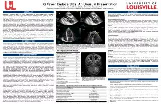
Case report Computed tomography (CT) of the petrous part of the temporal bone showed a left-sided 4-cm mastoid lytic process, extending upward toward the squamous temporalis, involving the outer and inner tables of the bone ..
Case report Axial CT scans of temporal bone showing the 4-cm lytic lesion of the left mastoid extending to the temporal squama
Case report • Bone curettage of diagnostic intent was performed. Microscopically, the product of this curettage showed a benign bone-forming tumor composed of a network of osseous trabeculae at variable stages of maturity within a highly vascular stroma . • These trabeculae were lined by one or several rows of active osteoblasts, with scattered associated osteoclasts. These findings were consistent withbenign OB. (H&E×100): trabeculae at varying stages of maturity. (H&E×200): trabeculae surrounded by two or more rows of active osteoblasts. European Annals of Otorhinolaryngology, Head and Neck diseases (2010) 127, 183—185
Case report • The patient was lost to follow-up and was seen again only in January 2009, with a lesion that appeared stable from a clinical and radiological points of view. • The patient under- went surgery in June 2009, with ablation of the tumor, which had extended to come in contact with the dural mater, with reaming of the healthy bone margins. • Histological examination of the gross specimen confirmed the diagnosis of OB. Postoperative recovery was uneventful and the patient was well without any sign of recurrence • 1 year after surgery.
Discussion • OB is a rare bone-forming benign tumor, accounting for 1% of • all bone tumors . In the head and neck, it often involves the • cervical vertebrae, the maxillary bones, or the frontal bone. • Location on the temporal bone is rare and on the mastoid process even rarer . To our knowledge, the case reported • herein is the 14th OB of the mastoid described in the literature .
Discussion • In its usual locations, OB is more frequent in male subjects aged between 10 and 30 years (90% of cases). • A clear female predominance has been noted in head and neck locations, particularly with the temporal bone . • Symptoms include tumefaction associated with continuous bone pain that are not relieved by minor antalgics, more rarely by tinnitus, reduced auditory acuity, or facial paralysis following involvement of the adjacent cranial nerves .
The radiological appearance of the tumor on CT scans is that of • a round or oval, well-demarcated lytic lesion, with varying • degrees of calcification . Discussion Axial CT scan of temporal bone showing a 4-cm lytic lesion of the left mastoid extending to the temporal squama , Axial bone-target computed tomographic scan image showing a bony destructive mass lesion with calcified component involving the jugular foramen, mastoid air cell, middle ear, and posterior part of the pyramid. Surgical Neurology 66 (2006) 534– 538
Discussion • The MRI findings of benign osteoblastoma are characterized by an hypointense or isointense mass on a T1-weighted image with homogenous or heterogeneous enhancement after the administration of gadolinium and by a hypointense mass on a T2-wieghted image MR scan images showing isointensity on a T1-weighted image (A), low intensity on a T2-weighted image (B), and remarkable enhancement on a gadolinium-enhanced T1-weighted image (C) in the left temporal bone. Surgical Neurology 66 (2006) 534– 538
Discussion • In some forms of aggressive OB, the radiological appearance is very misleading and can mimic osteosarcoma, especially in flat bones . • In cases of intracranial development, the radiological differential diagnosis with meningioma can be difficult.
Conclusion • OB of the temporal bone, and in particular, of the mastoid, is very rare, making its preoperative diagnosis very difficult. • It most often affects young females and presents as a slow growing painful lump.