Download
1 / 48
480 likes | 488 Views
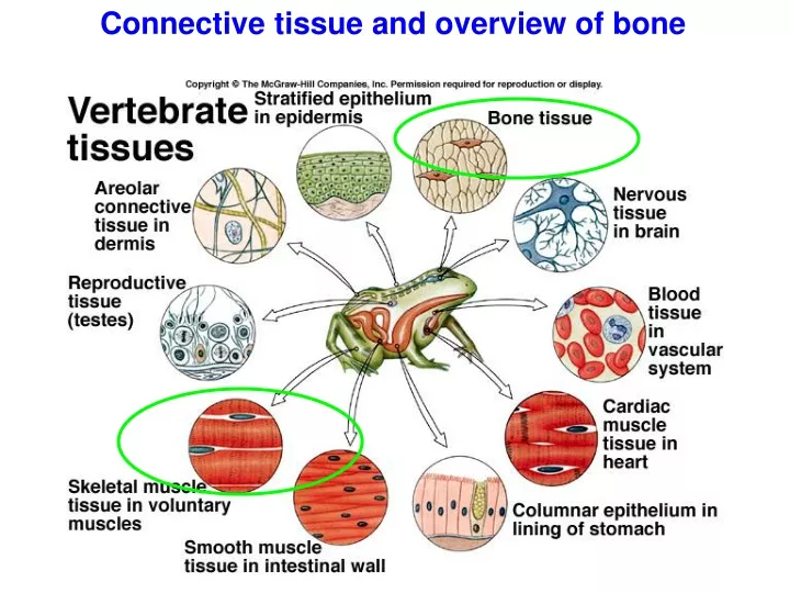
Connective tissue and overview of bone. Hyaline cartilage. Fibrous cartilage. Elastic cartilage. Bone shapes. Flat (parietal/skull). Short (carpels). Long (humerus). Irregular (vertebra). Axial Skeleton. Joints (=Articulations):

E N D
Hyaline cartilage Fibrous cartilage Elastic cartilage
Bone shapes Flat (parietal/skull) Short (carpels) Long (humerus) Irregular (vertebra)
Joints (=Articulations): Fibrous—connect bones but without movement (skull, pelvis) Cartilaginous—connect bones with cartilage, as in vertebrate and ribs Synovial—connect bones and allow movement
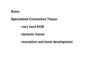
General types of bone Slow-growing, collagen regular Osteons form with canals for blood & lymph vessels, and nerves Fast-growing, collagen irregular
Intramembranous Bone Development • occurs at numerous sites simultaneously • see outer compact bone and inner, forming, spongy bone • mesenchyme, not cartilage, is replaced • Includes dermal bone (skull, pectoral girdle, integument), sesamoid bones (e.g., patella), perichondral and periosteal bone forming bone
Endochondral bone formation and ossification in a human fetus
Bone density scanning =dual-energy x-ray absorptiometry (DXA) Useful for tracking osteoporosis
Astronauts in space need to exercise too! Astronaut Peggy Whitson exercises during her stay aboard the International Space Station. Credit: NASA, Your Body in Space: Use it or Lose It Astronaut Charlie Hobaugh performing exercise on the iRED on the KC-135. Special exercise equipment is needed in the microgravity environment aboard the International Space Station. Regular weights are "weightless" in space; exercise equipment is designed to resist lifting and pulling and pushing so that astronauts can get a healthy workout and maintain their muscles and bones. Credit: NASA, About the Exercise Physiology Laboratory
The Muscular System Dr. Gunther van Hagens Body Worlds
X-section Muscle types Smooth involuntary muscle sagittal section Striated voluntary (skeletal) muscle 1. muscle fiber 2. nuclei Cardiac muscle 1. cardiac muscle cell 2. nuclei 3. Intercalated discs
Muscle cell (fiber) characteristics SmoothSkeletal Cardiac Not striated Striated Striated Spindle-shaped Cylindrical Cylindrical Not branched Not branched Branched Single central nucleus Multiple peripheral nuclei Single, central nucleus No discs No discs Intercalated discs Involuntary Voluntary Involuntary Slow Fast and slow Fast Contraction not inherent Contraction not inherent Contraction inherent Tonic fibers Twitch fibers
Many muscle fibers (cells) surrounded by endomysium skeletal muscle surrounded by epimysium Mitochondria in muscle cells bloodvessels muscle fascicle surrounded by perimysium nerve
Myofibril Muscle fiber (=cell)
Sarcomere: functional unit of a skeletal muscle: =made up of actin(thin filaments) and myosin(thick filaments) that interact during a contraction. Sliding filament http://www.youtube.com/watch?v=EdHzKYDxrKc
Sarcomere Building a myofibril = repeating modules (sarcomeres)
Building a muscle cell, or fiber =bundle of myofibrils
Figure 1 - Cut-away diagram of skeletal muscle (mammalian) showing several microfibrils and associated sarcoplasmic reticulum and the sarcolemma and t-tubular systems Figure 2 - A cartoon showing the sarcolemma, T-tubules and SR membranes and the junctional end feet (that span the gap between the t-tubular and SR membranes)
Check out animation of a muscle contraction: http://www.sci.sdsu.edu/movies/actin_myosin.html • Actin is enveloped in proteins (troponin and tropomyosin) • Without Ca++, tropomyosin blocks myosin binding site (myosin can’t access actin) • A motor neuron triggers wave of electrical signal to be sent to sarcolemma via transverse tubule • Electrical signal triggers Ca++ release from sarcoplasmic reticulum • Ca++ release allows myosin to bind to actin (forms cross-bridge) • When myosin molecule relaxes (after converting ATP to ADP) Another fabulous instructional video: http://www.youtube.com/watch?v=gJ309LfHQ3M
Muscle tension and the motor unit Depends on frequency of stimulation & number of fibers stimulated http://www.anatomybox.com/neuromuscular-junction/ Histological visualization of neuromuscular junctions in muscle. The motor neuron is depicted in green (anti-neurofilament immunohistochemistry) and the acetylcholine receptor clusters on the muscle fiber membrane in red (using fluorescently labeled alpha-bungarotoxin).
Muscle actions Motor pattern = any repetitive movement activated by the nervous system; involves coordinated efforts by different muscles e.g., coughing to clear throat uses 253 muscles! Synergistic muscles = work together to produce motion in same general direction (e.g., biceps brachii and brachialis to flex the forearm) Antagonistic muscles = produce opposing motions (e.g., biceps brachii and triceps brachii)
Muscle actions Flexor: bending about a joint Extensor: straighten a part Adductor: draw limb toward midline Abductor: move limb away from midline Levator: close jaw Depressor: open jaw Protractor: project away from base Retractor: move towards base
Muscle fiber types Type I = Slow oxidative (SO) Type IIa = Fast oxidative glycolytic (FOG) Type IIb = Fast glycolytic
cap mi cap cap cap From Ressel, 1996