Download
1 / 72
720 likes | 722 Views
NINE ABDOMINO-PELVIC REGIONS. Maintaining Homeostasis. The body communicates through nervous and endocrine systems consisting of 3 basic components 1) Receptor Detects a stimulus 2) Control center Analyzes information Determines appropriate response

E N D
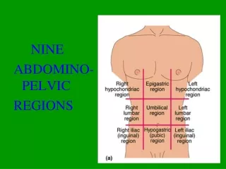
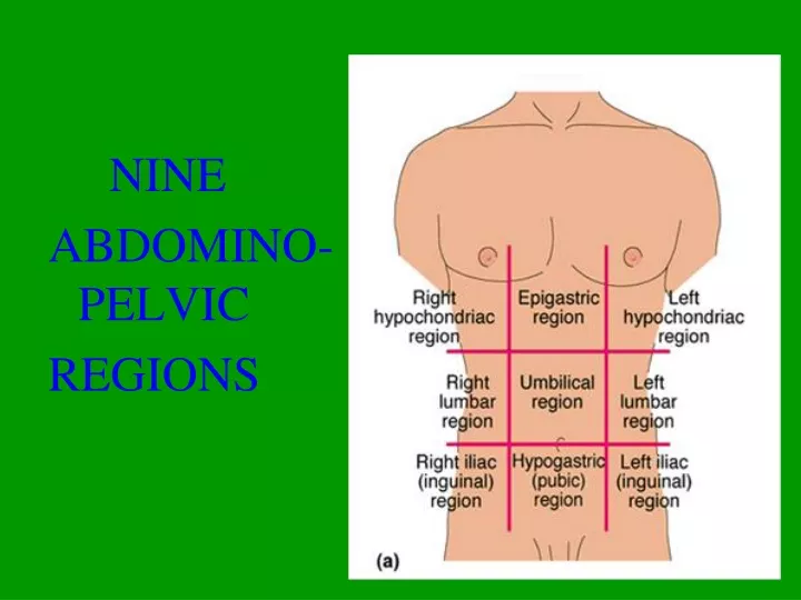
NINE ABDOMINO-PELVIC REGIONS
Maintaining Homeostasis • The body communicates through nervous and endocrine systems consisting of 3 basic components 1) Receptor • Detects a stimulus 2) Control center • Analyzes information • Determines appropriate response 3) Effector (Muscles or glands) • Responds to the stimulus
Trapezius Deltoid Pectoralis Major Bicep Rectus Abdominus Sartorius Quadriceps Anterior Tibialis Anterior Muscle Man
Trapezius Deltoid Rhomboid Tricep Latissimus Dorsi Gluteus Maximus IT Band Hamstring Gastrocnemius Posterior Muscle Man
Trapezius Rhomboid Deltoid Latissimus Dorsi The Back
Latissimus Dorsi Latissimus Dorsi
_________ Pectoralis Major ________ Serratus Anterior Chest Complex
Anterior Upper Leg 2. Psoas Major 6. Sartorius 9. Rectus Femoris 10. Vastus Medialis Oblique 11. Vastus Lateralis
1. Gluteus Medius 2. Gluteus Maximus 3. Tensor Fascia Lattae 4. IT Band Lateral Upper Leg
Posterior Upper Leg 1. Gluteus Medius 2. Gluteus Maximus 3. IT Band 4. Semitendiosis 5. Biceps Femoris 6. Semimembranosis
1. Gluteus Medius 2. Gluteus Maximus 3. Gluteus Minimus 4. Piriformis Posterior Muscles
Posterior Lower Leg 1. Gastrocnemius 2. Soleus 4. Achilles Tendon
Cranium Skull Facial bones Clavicle Thoracic cage (ribs and sternum) Scapula Sternum Rib Humerus Vertebra Vertebral column Radius Ulna Sacrum Carpals Phalanges Metacarpals Femur Patella Tibia Fibula Tarsals Metatarsals (a) Anterior view Phalanges Figure 7.1a
C1 Cervical curvature (concave) 7 vertebrae, C1–C7 Spinous process Transverse processes Thoracic curvature (convex) 12 vertebrae, T1–T12 Intervertebral discs Intervertebral foramen Lumbar curvature (concave) 5 vertebrae, L1–L5 Sacral curvature (convex) 5 fused vertebrae sacrum Coccyx 4 fused vertebrae Anterior view Right lateral view Figure 7.16
Movements at Synovial Joints • Gliding • Angular movements: • Flexion, extension, hyperextension • Abduction, adduction • Circumduction • Rotation • Medial and lateral rotation
Movements at Synovial Joints 4. Special movements • Supination, pronation • Dorsiflexion, plantar flexion of the foot • Inversion, eversion • Protraction, retraction • Elevation, depression • Opposition
Gliding Movements • One flat bone surface glides or slips over another similar surface • Examples: • Intercarpal joints • Intertarsal joints • Between articular processes of vertebrae
Gliding (a) Gliding movements at the wrist Figure 8.5a
Angular Movements Movements that occur along the sagittal plane: • Flexion—decreases the angle of the joint • Extension— increases the angle of the joint • Hyperextension—excessive extension beyond normal range of motion
Hyperextension Extension Flexion (b) Angular movements: flexion, extension, and hyperextension of the neck Figure 8.5b
Extension Hyperextension Flexion (c) Angular movements: flexion, extension, andhyperextension of the vertebral column Figure 8.5c
Flexion Extension Flexion Extension (d) Angular movements: flexion and extension at theshoulder and knee Figure 8.5d
Angular Movements Movements that occur along the frontal plane: • Abduction—movement away from the midline • Adduction—movement toward the midline • Circumduction—flexion + abduction + extension + adduction of a limb so as to describe a cone in space
Abduction Circumduction Adduction (e) Angular movements: abduction, adduction, andcircumduction of the upper limb at the shoulder Figure 8.5e