Download
1 / 43
430 likes | 518 Views
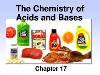
Acids, Bases, & the pH Scale. 2-17. Acids & Bases. Acids release protons (H + ) in a solution (proton donor) Bases lower H+ levels of a solution (proton acceptor). 2-18. pH. Is symbol for H+ concentration of a solution pH scale runs from 0 to 14 pH = log 1 [H + ]

E N D
Acids & Bases • Acids release protons (H+) in a solution (proton donor) • Bases lower H+ levels of a solution (proton acceptor) 2-18
pH • Is symbol for H+ concentration of a solution • pH scale runs from 0 to 14 • pH = log 1 [H+] • Pure H20 is neutral & has pH of 7 • Acids have a pH less than 7 (pH 0 - 7) • Bases have a pH greater than 7 (pH 7 - 14) 2-19
pH continued 2-20
Buffers • Are molecules that slow changes in pH by either combining with or releasing H+s • E.g. the bicarbonate buffer system in blood: H20 + C02 H2C03 H+ + HC03- • This buffers pH because reaction can go in either direction depending upon concentration of H+s 2-21
Blood pH • Normal range of pH is 7.35 – 7.45 • Maintained by buffering action • Acidosis occurs if pH < 7.35 • Alkalosis occurs if pH > 7.45 2-22
Organic Molecules 2-23
Organic Molecules continued • Are those that contain carbon & hydrogen • Carbon has 4 electrons in outer shell • Bonds covalently to fill outer shell with 8 electrons 2-24
Organic Molecules continued • In body, carbons are linked to form chains or rings • Serve as “backbone” to which more reactive functional groups are added Fig 2.9 2-25
Organic Molecules continued • Functional groups: • Carbonyl group forms ketones & aldehydes • Hydroxyl group forms alcohols • Carboxyl group forms organic acids (lactic & acetic acids) Fig 2.11 2-26
Stereoisomers • Contain same atoms arranged in same sequence • Differ in spatial orientation of a functional group • D-isomers are right-handed • L-isomers are left-handed • Biological enzymes act on only 1 of stereoisomers • E.g., enzymes of all cells can use only L-amino acids & D-sugars 2-27
Carbohydrates 2-28
Carbohydrates Fig 2.13 • Are organic molecules containing carbon, hydrogen & oxygen in ratio of CnH2n0n • Monosaccharides are simple sugars such as glucose, fructose, galactose 2-29
Carbohydrates continued • Disaccharides are 2 monosaccharides joined covalently • Include: • Sucrose (=glucose + fructose) or table sugar • Lactose (=glucose + galactose) or milk sugar • Maltose (=2 glucoses) or malt sugar 2-30
Carbohydrates continued • Polysaccharides are many monosaccharides linked together • Include starch & glycogen which are polymers of thousands of glucoses • Energy storage molecules • Allows organisms to store thousands of glucoses in 1 polysaccharide molecule which drastically reduces osmotic problems 2-31
Formation of Disaccharides • Occurs by splitting water out of 2 monosaccharides • An H+ & OH- are removed, producing H20 • Called dehydration or condensation 2-32
Digestion of Polysaccharides • Is reverse of dehydration synthesis • H20 is split, H+ added to one monosaccharide, OH- to other -- called hydrolysis • Polysaccharide hydrolyzed into disaccharides, then to monosaccharides 2-33
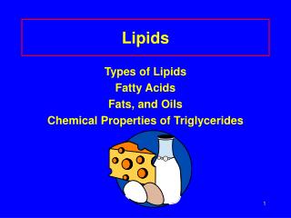
Lipids 2-34
Lipids • Are insoluble in polar solvents such as water • Hydrophobic • Consist primarily of hydrocarbon chains & rings 2-35
Lipids - Triglycerides • Formed by condensation of 1 glycerol & 3 fatty acids Fig 2.18 2-36
Lipids - Triglycerides continued • Are saturated if hydrocarbon chains of fatty acids are joined by single covalent bonds • Are unsaturated if there are double bonds within hydrocarbon chains 2-37
Lipids - Ketone Bodies • Hydrolysis of triglycerides releases free fatty acids • Which can be used for energy • Or converted in liver to ketone bodies • Which are acidic • High levels = ketosis • Ketoacidosis occurs when ketone bodies in blood lower pH Fig 2.19 2-38
Lipids - Phospholipids Lecithin • Are lipids that contain a phosphate group • Phosphate part is polar & hydrophilic • Lipid part is nonpolar & hydrophobic Fig 2.20 2-39
Lipids - Phospholipids micelle Fig 2.21 • Aggregate into micelles in water • Polar part interacts with water; nonpolar part is hidden in middle • Act as surfactants by reducing surface tension 2-40
Lipids - Steroids • Are nonpolar & insoluble in water • All have three 6-carbon rings joined to a 5-carbon ring • Cholesterol is precursor for steroid hormones • & is component of cell membranes Fig 2.22 2-41
Lipids - Prostaglandins • Are fatty acids with cyclic hydrocarbon group • Produced & active in most tissues • Serve many regulatory functions Fig 2.23 2-42
Proteins 2-43
Proteins - Amino Acids • Are made of long chains of amino acids • 20 different amino acids can be used • Amino acids contain an amino group (NH2) at one end; carboxyl group (COOH) at other end • Differences between amino acids are due to differences in functional groups (“R”) Fig 2.24 2-44
Proteins - Peptides • Are short chains of amino acids • Amino acids are linked by peptide bonds • Formed by dehydration reactions Fig 2.25 2-45
Proteins - Peptides • If <100 amino acids is called a polypeptide; >100 amino acids, is called a protein 2-46
Proteins - Structure • Can be described at 4 levels • Primary structure is its sequence of amino acids Fig 2.26a 2-47
Proteins - Structure continued • Secondary structure is caused by weak H bonding of amino acids • Results in alpha helix or beta pleated sheet shapes Fig 2.26b,c 2-48
Protein - Structure continued • Tertiary structure is caused by bending & folding of polypeptide chains to produce 3-dimensional shape • Formed & stabilized by weak bonds between functional groups • Not very stable; can be denatured by heat, pH Fig 2.26d 2-49
Protein - Structure continued • Figure 2.27 shows bonds responsible for tertiary structure Fig 2.27 2-50
Protein - Structure continued • Quaternary structure forms when a number of polypeptide chains are covalently joined Fig 2.26e 2-51
Protein - Structure continued • Many proteins are conjugated with other groups • Glycoproteins contain carbohydrates • Lipoproteins contain lipids • Others, like hemoglobin, contain a pigment 2-52
Nucleic Acids 2-53
Nucleic Acids Fig 2.29 • Include DNA & RNA • Are made of long chains of nucleotides • Which consist of a 5-carbon sugar, phosphate group, & nitrogenous base • Bases are pyrimidines (1 ring) or purines (2 rings) 2-54
Nucleic Acids - DNA • Contains genetic code • Its deoxyribose sugar (5C) is covalently bonded to 1 of 4 bases: • Guanine or adenine (purines) • Cytosine or thymine (pyrimidines) • Chain is formed by sugar of 1 nucleotide bonding to phosphate of another 2-55
Nucleic Acids - DNA continued • Each base can form hydrogen bonds with other bases • This hydrogen bonding holds 2 strands of DNA together 2-56
Nucleic Acids - DNA continued • The 2 strands of DNA twist to form a double helix • Number of purines = pyrimidines • Due to law of complementary base pairing • adenine pairs only with thymine; cytosine with guanine 2-57
Nucleic Acids - RNA • Consists of a long chain of nucleotides joined together by sugar-phosphate bonds • Its ribose sugar is bonded to 1 of 4 bases: • Guanine or adenine • Cytosine or uracil (replaces thymine) • Single-stranded 2-58
Nucleic Acids - RNA • 3 types of RNA are synthesized from DNA & allow it to direct activities of a cell: • Messenger RNA - mRNA • Transfer RNA - tRNA • Ribosomal RNA - rRNA 2-59