Download
1 / 42
420 likes | 429 Views
Plenary session 3: MALDI-TOF progress in clinical mycology. MALDI identification and more of moulds Maurizio Sanguinetti Institute of Microbiology Fondazione Policlinico Universitario «A. Gemelli» - Rome - Italy.

E N D
Plenary session 3: MALDI-TOF progress in clinical mycology MALDI identification and more of mouldsMaurizio SanguinettiInstitute of Microbiology Fondazione Policlinico Universitario «A. Gemelli» - Rome - Italy
Invasive mould infections continue to occur among immunocompromised patients, particularly those receiving haematopoietic stem cell or organ-solid transplantation. These infections are associated with high mortality rates. Beyond Aspergillus species, other moulds (or filamentous fungi) such as Fusarium and Scedosporium species and Mucoromycotina (Rhizopus, Mucor and Lichtheimia corymbifera) have emerged as causes of life-threatening infections.
While A. fumigatus sensu stricto represents by far the leading human pathogen, some other species belonging to the A. fumigatus sensu latu complex (i.e. section Fumigati) have been recognized as occasional causes of invasive aspergillosis in 3 to 6% of cases [Lamoth, Front Microbiol. 2016] Prevalence of Aspergillus spp. of section Fumigati other than Aspergillus fumigatus in clinical specimens aTeleomorph: Neosartorya pseudofischeri
Case reports of IA attributed to Aspergillus spp. of section Fumigati other than A. fumigatus From: Lamoth, Front Microbiol. 2016
Outbreaks and unusual sources of emerging moulds From: Miceli & Lee, Mycoses 2011
Susceptibilities of selected species belonging to the genus Aspergillus
Conventional Mould ID Therefore, our ability to diagnose fungal infections with conventional methods has been complicated, in recent years, by the evolving and widening “spectrum” of human fungal pathogens At present, conventional laboratory identification of moulds relies on the analysis of colony morphology and microscopic characteristics, BUT such phenotypic ID is time- and labour-consuming and highly subjective
Identification of filamentous fungi remains difficult Even DNA-sequence based classification of moulds has several limitations Biological complexity Changes in classification due to the introduction of nucleic-acid based techniques
Conventional Mould ID The literature constantly provides examples of morphological minutia to describe new species, as well as examples of “cryptic” species (i.e. species morphologically indistinguishable but separable using molecular methods) If fungal cultures remain integral in modern mycology laboratories, identification of the colony-cultured mould can be accelerated by use of the new proteomic technology, namely MALDI-TOF MS
The success of MALDI-TOF MS: key features Mass spectra are species-specific fingerprints, highly reproducible, and only minimally influenced by growth conditions A single sample is analyzed within minutes, with a time gain of one or more days compared with the conventional identification techniques. Although bacteria are traditionally identified in 1 day, longer times are required for molds (2–4 weeks) This comparative “time quantitation” is needed to perceive the dramatic impact that MALDI-TOF MS has on the workflow in a clinical microbiology laboratory
A generated MALDI-TOF mass spectrum never matches a reference “main spectrum” (MSP) with absolute identity. The result of mass spectral comparison, for a given microbial sample, is expressed by a value which represents the degree of similarity against a list of species matches • The Biotyper software generates a “score value” ranging from 0 to 3, with a score ≥2 as a criterion of acceptance at the species-level identification and a score ≥1.7 at the genus-level identification • The SARAMIS software generates a “confidence value” expressed as percentage of identity with the MSP, with >90% recommended for species-level identification and >70% for genus-level identification • The Andromas software reports percentages as identification criteria 70% is +/- comparable to a Bruker log(score) of 1.7 From: Posteraro et al. Expert Rev Proteomics. 2013
MALDI-TOF Identification of Filamentous Fungi From: Posteraro et al. Expert Rev Proteomics. 2013 • Acquisition of MALDI-TOF MS in the clinical mycology laboratory moved slowly with respect to the bacteriology setting. This, perhaps, because of: • Intrinsic difficulty of studying fungi as a whole due to their biological complexity • Coexistence of different (hyphal or conidial) fungal phenotypes, also in the same organism isolated from a mycological culture • Very few spectra currently included in the database of commercially available devices
Unlike for yeasts, MALDI-TOF MS-based identification of moulds remains reserved for a small number of highly specialized laboratories Mould identification using this technology still requires the establishment of comprehensive databases covering the vast diversity of fungi involved in human pathologies Generally, better identification scores are obtainable from extracted-protein suspensions rather than from direct colony deposition MALDI-TOF Identification of Filamentous Fungi
Key issues for routine MALDI-TOF MS analysis of moulds Sample preparation Reference database Interpretive cut-offs
Sample preparation Intact cell (IC), also termed “whole cell”, mass spectrometry (ICMS) approach — may be hindered by the presence of a robust cell wall in fungi Cell lysis (CL) approach — includes an extraction step with acetonitrile and therefore referred as “complete extraction” The IC method is recommended for use with the Andromas and SARAMIS systems, whereas CL is recommended for use with the Bruker Biotyper and Vitek MS systems — BioMérieux moved from an IC approach toward a CL approach
Sample preparation To minimize the effect of growth conditions on the production of a uniform mycelium by fungi, in 2012, Bruker Daltonics launched an additional spectral library, Filamentous Fungi Library 1.0, that was constructed by using 24-h-old (48) liquid cultures for complete protein extraction. This library is separate from the general (Bruker Biotyper) library and represents the first commercial database by Bruker Daltonics for the identification of molds grown in liquid media.
Using Bruker Biotyper software in conjunction with Filamentous Fungi Library 1.0, but without liquid culture-based sample preparation, a confident MALDI-TOF MS identification (cutoff value of 1.7) was shown for 91.6% (44/48) of mold isolates tested by Riat et al. (Int J Infect Dis 35:43– 45). Sleiman et al. used mechanical lysis followed by complete extraction of molds grown on solid media and an Australian database for the identification of 28 Aspergillus, Scedosporium, and Fusarium species (J Clin Microbiol 54: 2182–2186). This in-house database, combined with Bruker Filamentous Fungi Library 1.0, outperforms the Bruker library alone in terms of rates of correct species identification (93% versus 69% for Aspergillus, 84% versus 42% for Fusarium, and 94% versus 18% for Scedosporium, respectively). Sample preparation
Sample preparation In a previous study (Clin Microbiol Infect 2012, 18:475–484), we pre-treated the sample simply by mixing fungal material (mycelium and/or conidia) with distilled water. This is consistent with attempts, also done by several research groups, to simplify preanalytical processes in order to avoid the clinical (i.e. delayed turnaround time) and laboratory (i.e. prolonged working time) consequences of using liquid mold cultures. Of course, the risk of aerosols and contamination is greater with extensive handling of moulds grown on agar than with moulds grown in broth. However, the use of a biosafety cabinet during sample processing can help to prevent potential infections of personnel working near a mycology laboratory.
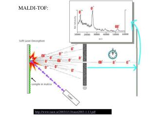
Work-flow for mouldidentification by MALDI-TOF MS Unknownmould MALDI-TOF MS processing Absolute ethanol HCCA matrix MALDI-TOF spectrum profile acqusition H2O Speciesidentification Match to fungi database YES Log(score)≥2.0? Data entry in fungi database Molecularidentification NO
Reference database The reference database provided with each commercial MALDI-TOF MS platform may not be sufficient for routine analyses (shown later). The most clinically comprehensive mould databases, developed recently by Lau et al. (J Clin Microbiol 2013, 51:828–834), Gautier et al. (Clin Microbiol Infect 2014, 20:1366–1371), and Becker et al. (Med Mycol 2014, 52:826–834), contained 152, 347 and 472 fungal species, respectively. In the meantime, a homemade library for MALDI-TOF MS identification of dermatophytes was used in conjunction with the Bruker Biotyper library (version 3.0), which contained 3,995 unique reference spectra (Theel et al., J Clin Microbiol 2011, 49:4067–4071). In another study, using the SARAMIS system, SuperSpectra were created for MALDI-TOF MS identification of dermatophytes (De Respinis et al., Med Mycol 2013, 51:514–521).
Head-to-head comparison of the Bruker Biotyper and Vitek MS systems A large collection of microorganisms, including filamentous fungi, at a public health reference laboratory in Quebec (Canada) were studied. • Only 17 of 71 isolates tested were referenced in both systems’ databases, with 43 being present in the Bruker Biotyper database and 20 being present in the Vitek MS database. • Thus, when present in their respective databases, 76.7% (33/43) and 50.0% (10/20) of isolates could be correctly identified (to both the species and genus levels) with the Bruker Biotyper and Vitek MS systems, respectively. Only one misidentification was obtained (1 Fusarium proliferatum isolate misidentified as Fusarium oxysporum) with the Vitek MS system, whereas there was no misidentification with the Bruker Biotyper system. • When absent from their respective databases, 12 misidentifications (mainly to the genus level) were obtained with the Vitek MS system, as opposed to none with the Bruker Biotyper system.
Based on a procedure proposed previously by Cassagne et al. (PLoS One 2011, 6:e28425) but using only in-house databases for moulds (in combination with the Bruker Biotyper library), studies have shown that MALDI-TOF MS could be reliable for the identification of any species of clinically relevant mould, including the “cryptic”Aspergillus species. However, these studies applied a lower species cut-off (mostly 1.7 [value accepted for genus assignment]) to increase identification rates. Interpretive cut-offs
Shown are examples that specifically address key points such as the accurate identification of cryptic or closely related species, identification of nonsporulating or poorly sporulating organisms (e.g. dermatophytes), and rapid identification of clinically significant organisms such as dimorphs or emerging fungi. Remarkably, all these studies used the ethanol-formic acid extraction method before MALDI-TOF MS analysis of their mould samples.
Examples of mould identification by MALDI-TOF MS Aspergillus Species Fusarium Species Rhizopus-Lichtheimia Species
Using the BrukerBiotyper database (OC version 3.1; updated January 2016) amended with an in-house fungal database to expand the number of identifiable Aspergillusspecies, Masih et al. reported successful MALDI-TOF MS identification results (score value of 2.0) for 97.7% of 45 clinically significant Aspergillusisolates. The isolates belonged to 23 rare Aspergillusspecies (only 8 of which are present in the current BrukerBiotyper database) that were enclosed in 12 sections (mainly sections Circumdati, Nidulantes, Flavi, Terrei, Versicolores, Aspergillus and Nigri). Interestingly, two cryptic Aspergillusspecies, A. pallidofulvus(section Circumdati) and A. aegyptiacus(section Usti), were isolated for the first time from clinical specimens.
Triest et al. developed a user-friendly identification approach relying upon an in-house database, which was constructed with spectra from 289 validated strains belonging to 40 Fusarium species. For 19 species represented by more than one strain, it was found that by using MALDI Biotyper 3.0 software and a score value of 2.0 as the cut-off for identification, 82.8% of MALDI-TOF MS-based identifications were correct at the species level, 3% were correct at the species complex level, and the remaining 14.2% were not reliable. Interestingly, the success rate of correct identifications increased to even 97.0% when some Fusarium species complexes were taken as a whole; thus, MALDI-TOF MS identification would have failed with only 4 Fusarium (1 F. incarnatum, 1 F. equiseti, 1 F. sporotrichioides, and 1 F. sacchari) strains in the study. Representation of the distance matrix dendrogram of Fusarium reference spectra. The different species clades are shown. Most strains of a same species fall into a single clade or closely related clades.
MALDI-TOF MS can be used to clearly discriminate Lichtheimia species from other pathogenic species of the Mucorales. The reliability and robustness of the MALDI-TOF-based identification are evidenced by the ability to discriminate between clinically relevant (Lichtheimia corymbifera, L. ramosa, and L. ornata) and irrelevant (L. hyalospora and L. sphaerocystis) species. In total, all 34 strains were unequivocally identified by MALDI-TOF MS to the generic level, 32 out of 34 of the Lichtheimia isolates were identified accurately with score values of >2 (probable species identification), and 25 of 34 isolates were identified to the species level with score values of >2.3 (highly probable species identification). The MALDI-TOF MS-based method reported here was found to be reproducible and accurate, with low consumable costs and minimal preparation time.
Mould identification byMALDI-TOF MS: Is it really a history of success Over the last 5 years, accumulated experience clearly shows that MALDI-TOF MS holds promise as an accurate mould identification tool, particularly with common filamentous fungal pathogens. The turnaround time has been reduced with the use of MALDI-TOF MS instruments in the clinical laboratory routine, but these instruments continue to rely on fungal cultures. A major limitation of MALDI-TOF MS for mould identification is still the breadth of commercially available databases. Hence the need for expanded databases, which is apparent, although not exclusively, for rare, emerging or endemic mycosis agents. Molecular methods (e.g. DNA sequencing) are currently the gold standard for the identification of fungi to the species level. • However, it is worth noting that Aspergillus flavus, the second leading cause of human aspergillosis, is not separable from Aspergillus oryzae by means of molecular biology techniques, whereas the closely related species Trichophyton mentagrophytes and Trichophyton interdigitale are not separable by means of ITS sequence analysis. • In contrast, MALDI-TOF MS seems to be more powerful for discrimination between these species, as well as to distinguish clinically relevant from irrelevant species of Lichtheimia.
This study assessed an online identification application based on original algorithms and an extensive in-house reference database comprising 11,851 spectra (938 fungal species and 246 fungal genera). • Validation criteria were established using an initial panel of 422 molds, including dermatophytes, previously identified via DNA sequencing (126 species). • The application was further assessed using a separate panel of 501 cultured clinical isolates (88 mold taxa including dermatophytes) derived from five hospital laboratories. • A total of 438 (87.35%) isolates were correctly identified at the species level, while 26 (5.22%) were assigned to the correct genus but the wrong species and 37 (7.43%) were not identified, since the defined threshold of 20 was not reached.
The use of the Bruker Daltonics database included in the MALDI Biotyper software resulted in a much higher rate of unidentified isolates (39.76 and 74.30% using the score thresholds 1.7 and 2.0, respectively). • Moreover, the identification delay of the online application remained compatible with real-time online queries (0.15 s per spectrum), and the application was faster than identifications using the MALDI Biotyper software. • This is the first study to assess an online identification system based on MALDI-TOF spectrum analysis and this approach to identify molds, including dermatophytes, for which diversity is insufficiently represented in commercial databases. • This free-accessapplication is available to medical mycologists to improve fungal identification.
Extended use of MALDI-TOF MS for moulds Closely related species differentiation Antifungal susceptibility testing
Cluster analysis of MALDI-TOF spectra of selected reference strains and challenge isolates (CH15, CH25 and CH51) identified as Aspergillus section Flavi species As several A. flavus isolates are known to be non(afla)toxigenic (and more similar to A. oryzae than to other A. flavusisolates), our findings raise the possibility of using this approach for discriminating toxigenic from atoxigenicA. flavusstrains From: De Carolis E, et al. Clin Microbiol Infect 2013;13:788–799
FUSARIUM-ID database SEQUENCE TYPE Mass spectrometry-baseddendrogram VS MLST The strainswereclustered in separate groupsaccordingtotheirphylogeneticspeciesdesignation • Each MSP is compared with the other in a matrix of cross-wise identification values. • The matrix is used to calculate the distance values for each pair • Based on the protein mass patterns, strains can be clustered hierarchically
We present the description and taxonomy of a new taxon, Fusarium ficicrescens sp. nov., collected from contaminated fig fruits in Iran. The species was studied by multilocus sequence analysis, amplified fragment length polymorphism (AFLP), matrix-assisted laser desorption ionization time-of-flight mass spectrometry (MALDI-TOF MS) and phenotypic characters. Phylogenetic analysis showed that the fungus is closely related to Fusarium lactis, Fusarium ramigenum, and Fusarium napiforme, known plant pathogens, mycotoxin producers, and occasionally occurring multidrug resistant opportunists. Two separate clusters were visualized by using MALDI-TOF MS, one comprising three strains including the type strain of F. andiyazi, and another with strains CBS 125177, CBS 125178, and CBS 125181. This indicates that MALDI-TOF MS has a potential to distinguish closely related and morphologically identical Fusarium species. The MALDI-TOF MS data are used as an additional parameter in the polyphasic classification of this cryptic species.
MIC determination Microdilution methods E-Test Disk diffusion It could take 48 h End-point subjective SUSCEPTIBILITY DETERMINATION New generation of susceptibility tests MASS SPECTROMETRY MEC ?
We developed a MALDI-TOF MS-based assay for testing antifungal susceptibilities of Candida and Aspergillusspecies to the echinocandinscaspofungin, by relying on the proteome changes which are detectable after a 15-h exposure of fungal cells to serial drug concentrations
By means of a composite correlation index (CCI)-based approach, the method reliably and accurately allows to determine the minimal profile change concentration (MPCC), an endpoint value that is an alternative to the classical MIC (Marinach et al. 2009) To date, while the endpoint readings achievable with MALDI-TOF MS represent a slight time-saving (15 h versus 24 h) over the CLSI/EUCAST method with respect to Candida species, MALDI-TOF MS has great advantage of eliminating subjective read-outs which occur with the CLSI (and EUCAST) method when filamentous fungi, such as Aspergillus species, are tested Using a panel of wild-type and fks mutant isolates of Candida (n = 34) and Aspergillus (n = 10) species, a full essential agreement between the values of MPCC and MIC (or MEC) was found for 100% of the isolates tested According to the MEC values, MPCC values of 0.5 and 0.25 μg/ml were able to capture, respectively, all of clinical A. fumigatus and Aspergillus flavus tested by us
“While the use of MALDI-TOF MS technology allowed for accurate identification of isolates as WT or non-WT, the current methodology did not save any time compared to traditional BMD assays. While the abbreviated MALDI-TOF MS method, which requires only 3 drug concentrations, may potentially reduce the amount of work required for setup, it still requires 30 to 48 h of incubation prior to analysis to allow time for sufficient growth for accurate differentiation between WT and non-WT strains”
CONCLUSIONS MALDI-TOF MS has contributed to improving the laboratory diagnosis of infections by filamentous fungi in terms of rapidity and accuracy of identification in the last few years. Future studies are required to estimate the real impact of MALDI-TOF MS identification results on the clinical and therapeutic management of mould diseases. Additional applications of MALDI-TOF MS-based diagnosis of fungal disease (i.e. antifungal susceptibility/resistance detection and fungal strain typing) need to be potentiated in the near future. In summary, progresses in these areas will aid in enhancing and diversifying the clinical diagnostic usefulness of MALDI-TOF MS.
Istituto di Microbiologia, Università Cattolica del Sacro Cuore, Roma, Italy E. De Carolis, A. Vella, R. Torelli, L. Vaccaro Istituto di Sanità Pubblica, Università Cattolica del Sacro Cuore, Roma, Italy B. Posteraro Public Health Research Institute, New Jersey Medical School, Rutgers, The State University of New Jersey, USA David S. Perlin Innsbruck Medical University, Innsbruck, Austria C. Lass-Flörl Dipartimento di Biotecnologie Cellulari ed Ematologia, Università La Sapienza di Roma, Italy C. Girmenia, C. Colozza Dipartimento Sanità pubblica Microbiologia e Virologia, Università degli Studi di Milano, Italy M. Cogliati, A. M. Tortorano