Download
1 / 48
540 likes | 1.14k Views
Neonatal Jaundice: Indirect Hyperbilirubinemia. Objectives. Identify risk factors for severe hyperbilirubinemia Understand the relationship between hyperbilirubinemia and the risk for neurologic or developmental injury

E N D
Objectives • Identify risk factors for severe hyperbilirubinemia • Understand the relationship between hyperbilirubinemia and the risk for neurologic or developmental injury • Discuss ways to screen for infants who might develop severe hyperbilirubinemia • Discuss guidelines for treatment
Epidemiology: Increased risk for neonatal jaundice • Infant Factors • Blood group incompatibilities: Rh, ABO, others • Hemolysis (non-isoimmune): infection, drugs, T-antigen exposure, coagulopathy, RBC enzyme deficiencies (G6PD, PK, HK), RBC structural defects (spherocytosis, elliptocytosis) • Hemorrhage: cephalohematomas, intracranial bleeding, bruising • Infection: sepsis, UTI • Endocrine: hypothyroidism, adrenal insufficiency
Epidemiology: Increased risk for neonatal jaundice • Infant Factors • Prematurity • Male • Polycythemia • Breast feeding vs. formula feeding • Caloric deprivation, postnatal weight loss • increased enterohepatic circulation • Delayed passage of meconium
Epidemiology: Increased risk for neonatal jaundice • Race: • Increased production: East Asian, Native American • G6PD: Greek, East Asian, African • Genetic: • History of sibling with jaundice • G6PD, hexokinase, pyruvate kinase deficiency • Gilbert’s syndrome, Crigler Najjar Syndrome • Spherocytosis, Elliptocytosis
Epidemiology: Increased risk for neonatal jaundice • Maternal diabetes mellitus: • Increased bilirubin production rate • Correlation with macrosomia and polycythemia • Elevated beta-glucuronidase in breastmilk • Maternal drugs: • epidural anesthesia (bupivacaine) • oxytocin • Delayed cord clamping
Epidemiology: Increased risk for neonatal jaundice • Environmental factors: • Phenolic detergents • Naphthalene (moth balls) • Short hospital stay • Failure to detect significant jaundice • Failure to establish breastfeeding
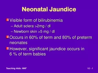
What is a normal “physiologic” serum bilirubin? • Dennery et al. NEJM 2001: average peak bilirubin in term newborn, 5-6 mg/dL • Breast fed infants are on average about 2 mg/dL higher than bottle fed infants in the first days of life. • Racial differences • Greek, Asian, Navajo reach higher peaks
How should non-physiologic jaundice be defined? • Collaborative Perinatal Project (1955-61) and Maisels (1986): upper limit of physiologic jaundice (95%) 12.9 mg/dL • Kaiser (1997): 95% = 17.5 mg/dL • Multicentered international study (Natus, 1998): 95% = 15.5 mg/dL, 2 SD = 17 mg/dL at 96 hours • Bhutani. Pediatrics 1999; 103:6 • Post discharge: 95th percentile 17.5 mg/dL • predictive curves for severe hyperbilirubinemia
JCAHO Sentinel Alert: April 2001 Root causes for re-admission for hyperbilirubinemia identified • Unreliability of visual assessment of jaundice • Failure to measure bilirubin before discharge or in an infant with visible jaundice in the first 24 hours • Early discharge: especially <38 weeks GA infant • Failure to provide early f/u assessment post- discharge • Failure to provide lactation support, information to parents about jaundice or poor feeding • Failure to treat appropriately
Strategies to prevent severe jaundice • Pre-discharge assessment (transcutaneous bilimeter or serum bilirubin) with use of Bhutani nomogram to predict risk • Standardized policies for screening • Follow-up of all newborns in 24-48 hr • Informational materials for parents about jaundice • Lactation support • Optimal application of phototherapy
Bhutani: hour specific serum bilirubin. Pediatrics 1999;103:6-14
Predictive nomograms for severe hyperbilirubinemia: Bhutani 1991 • What is the risk for subsequent “severe hyperbilirubinemia” (i.e. bilirubin level in the high risk zone, 95th%)? • > 95th %: 39.5% • 75-95th %: 21.6% • 40-75th %: 11.6% • < 40th %: virtually 0
Bilirubin follow-up policy • Compare serum bilirubin or transcutaneous bilirubin to Bhutani curves • > 95th%: repeat serum bilirubin in 24-48 hours • 75-95th%: repeat serum bilirubin in 24-48 hours • 40-75th%: if risk factors present, serum bilirubin in 24-48 hours • < 40th%: no follow-up needed
Bilirubin injury to the brain • Bilirubin encephalopathy: • Acute reversible changes • Acute irreversible changes • Kernicterus (yellow staining of the brain) • Neurodevelopmental sequelae • Clinical correlations • Epidemiologic studies
Clinical features of acute bilirubin encephalopathy • Acute form: • Early Phase 1 (1-2 days): poor suck, stupor, hypotonia, seizures • Intermediate Phase 2 (mid 1st week): hypertonia of extensor muscles, irritability, retrocollis-opisthotonus, fever • Advanced Phase 3 (after 1st week): irreversible CNS damage, retrocollis-opisthotonus, hypertonia, shrill cry, seizures, coma, apnea, death
Clinical features of kernicterus • Chronic form: • First year: hypertonia, active DTRs, obligatory tonic neck reflexes, delayed motor skills • > 1 year: movement disorders (choreoathetosis, ballismus, tremor), paralysis of upward gaze, hearing loss, mental retardation
Pathology of kernicterus • Orth: described bilirubin pigmentation of the brain in infants with severe jaundice in 1875 • Kernicterus: German word meaning jaundice of the nuclei • Term was coined by Christian Schmorl in 1904 • Yellow staining of the brain (basal ganglia) • Neuronal swelling • Death of neurons
Pathophysiology of bilirubin encephalopathy • Bilirubin monoanion binds to membrane • Causes changes in membrane characteristics • May affect membrane permeability • P-glycoprotein (PGP): ATP mediated transport of bilirubin across membranes and out of the cell • Activity low in immature animal • Can be inhibited by drugs: e.g., ceftriaxone • Membrane associated bilirubin oxidizing enzyme in the brain: activity low in immature animal
Pathophysiology of bilirubin encephalopathy • Blood brain barrier • Hyperosmolarity opens the barrier • Hypercarbia increases bilirubin deposition in the brain • Bilirubin binding to albumin: 1:1 at the first binding site • Displacement of bilirubin from albumin: sulfa drugs, benzyl alcohol, FFA, ceftriaxone
Cellular mechanisms of bilirubin toxicity • Binding to cellular membranes • Decreased Na-K exchange • Cellular accumulation of water • Axonal swelling • Lowering of membrane potentials, decreased action potential • Decreased amplitude and longer intervals in auditory response
Clinical factors which increase the risk for kernicterus or bilirubin encephalopathy • Displacement of bilirubin from albumin • Hyperosmolarity • Hypoxemia, hyperoxemia • Asphyxia • Hypercarbia • Acidosis • Sepsis • Hemolysis • Prematurity
Astute Observation from a Nurse • Sister J. Ward, Charge Nurse Premature Baby Unit, Rochford Hospital, Essex England 1957 • Skin of jaundiced infants bleached on exposure to sunlight, unexposed skin does not
The Science of Phototherapy • Bilirubin is a yellow pigment, absorbs blue light spectrum • Conversion of bilirubin into lumirubin, a water soluble compound • Elimination by the GI tract and kidney
Can You “Overdose” With Phototherapy? • “With existing equipment there is no such thing as an overdose of phototherapy” (Maisels2001) • The saturation point (where higher irradiance levels don’t matter) is not known
Phototherapy devices • White fluorescent tubes • Broad spectrum light exposure • Blue fluorescent tubes • Blue light is more effective • Blue LED lights (NeoBlue) • Halogen lamps • More compact, bulbs are hot and can burn if too close • Fiber optic blankets • small area of exposure
How Fast Can the Bilirubin Decline? • 6-20% decrease in 24 hours-”standard phototherapy” • 32% decrease in 18 hours- fiberoptic + bluelights • 43% decrease in 24 hours- blue lights above and below
Fluorescent Phototherapy Lights • Fluorescent lights cover more skin surface • Deliver higher intensity without heating • White lights effective, blue lights most effective • Bulbs lose intensity long before they “burn out”
Halogen Spotlight Phototherapy • Halogen spotlights heat skin if closer than 55cm • Cannot deliver higher “doses” of phototherapy • Bulbs burn out • Preferred by staff • More compact, easier to use in NICUs
Halogen Photometer Reading • “Double” halogen lights • Only able to generate 10 microwatts/cm2/nm • Very low “dose” of phototherapy
Fiberoptic Phototherapy • Light from tungsten-halogen bulb through fiberoptic cable • Less effective than conventional phototherapy • Should not be used in VLBW infants, potential for skin injury
Factors that determine dose and effectiveness of phototherapy • Spectrum of light (blue is best) • Irradiance of light source • power output of the lamp • Design of phototherapy device • does it expose the maximal amount of skin? • Surface area exposed to light • Distance of infant from light
Acute management of severe hyperbilirubinemia • Phototherapy with fluorescent or LED blue lights: maximal surface exposure and dose • Correct dehydration, acidosis (respiratory and metabolic), and hypotension • Correct hypoalbuminemia (1 g/dL of albumin binds 8.3 mg/dL bilirubin): augments removal of bilirubin with exchange transfusion • Reduce enterohepatic circulation of bilirubin: stop breast milk feedings, use formula feedings • PO charcoal and agar reported, but not commonly used
Acute management of severe hyperbilirubinemia • Avoid drugs which displace bilirubin from albumin or affect P glycoprotein • Avoid use of hyperosmolar drugs or infusions • Inhibitors of heme oxygenase (protoporphyrins): • Reduces bilirubin production • Sn and Zn protoporphyrins reported to be useful, but not yet FDA approved • Extra-corporeal removal of bilirubin: • theoretical possible • extracorporeal charcoal binding used in Russia
Recommendations for treatment of hyperbilirubinemia (AAP practice guideline) Age Consider Exchange if Exchange* (hr) phototherapy phototherapy photoRx fails# transfusion 25-48 > 12 > 15 > 20 > 25 48-72 > 15 > 18 > 25 > 30 >72 > 17 > 20 > 25 > 30 #Phototherapy should result in a decline 1-2 mg/dL of total bilirubin within 4-6 hour, should continue to fall and remain below exchange transfusion levels. *Intensive phototherapy, prepare for exchange, exchange if bilirubin does not fall below exchange transfusion levels. Adapted from Pediatrics 1994;94:558
Exchange transfusion: criteria • Term: > 30 mg/dL > 25 mg/dL, failed trial phototherapy • 35-36 weeks: > 25 mg/dL • 30-34 weeks: > 20 mg/dL • < 30 weeks: 15-20 mg/dL • Reduce exchange level 3-5 mg/dL for seriously ill infants: sepsis, acidosis, respiratory failure • Acute symptoms of bilirubin encephalopathy
Exchange Transfusion • ABO type-specific Rh negative blood in cases with Rh incompatibility • Type O Rh-specific cells in cases with cases with ABO incompatibility • Whole blood diluted with FFP to Hct of 50-55%. • Fresh blood < 24 hours old preferred. • Double volume exchange 160ml/kg
Technique for Exchange Transfusion • Withdrawal thru UA catheter with simultaneous infusion thru UVC catheter • 5-to 20-ml increments of warmed blood • Agitate blood every 10-15 minutes so cells don’t settle. • Initial sample sent for bilirubin, Hct, lytes, calcium, cultures
Things to Remember • Monitor ECG, BP, and temperature during procedure • Measure ABG at beginning, middle, and end of procedure. • Measure glucose at 10, 30, 60 minutes post procedure. • Measure calcium after each 100 ml of blood. • Warming blood > 37 degrees causes hemolysis
Bilirubin After Double Volume Exchange • Serum bilirubin is 45% to 60% of preexchange level
Infant Hypothermia Hyperkalemia Thrombocytopenia Low Ca++ and Mg++ Reactive hypoglycemia Action Warm donor blood Use fresh blood, monitor ECG Transfuse platelets at end if < 75K Give CaGluconate 100mg/kg/d IV glucose 5mg/kg/min 10-30 minutes after end of exchange Potential complications
Followup issues for hyperbilirubinemia • Hearing screen • “Rebound” bilirubin • AAP guideline: repeat bilirubin level not indicated in healthy term infants • useful in premature infants, hemolysis (isoimmunization, G6PD) • Infants with bilirubin encephalopathy • neurodevelopmental followup • hearing screen