Download
1 / 32
370 likes | 439 Views
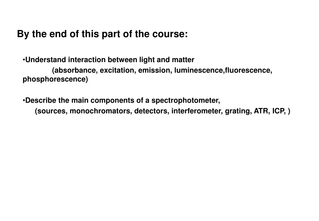
By the end of this part of the course:. Understand interaction between light and matter (absorbance, excitation, emission, luminescence,fluorescence, phosphorescence) Describe the main components of a spectrophotometer,

E N D
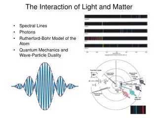
By the end of this part of the course: • Understand interaction between light and matter (absorbance, excitation, emission, luminescence,fluorescence, phosphorescence) • Describe the main components of a spectrophotometer, (sources, monochromators, detectors, interferometer, grating, ATR, ICP, )
And of course, the relationship between energy and frequency: E = hn = hc/l = hc n h = Planck’s constant (6.626 x 10⁻³⁴ J s) n = wavenumber (most common units = cm-1) ~ ~ Review on properties of light:photon Light is energy in the form of electromagenetic field Wavelength (l): Crest-to-crest distance between waves Frequency (n): Number of complete oscillations that the wave makes each second units: number of oscillations/sec or s-1 or Hertz |(Hz) Light travelling speed: in other media: c/n (n = refractive index, generally >1) in a vacuum: c=2.998 x 10⁸ m s-1 (n=1 exactly, in air n=1.0002926) c/n= nl Therefore: Energy is inversely proportional to wavelength but proportional to wavenumber
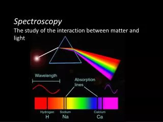
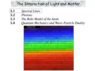
Frequency Scanning Techniques: a few definitions Emission method: source of light is sample Absorption method: intensities of a source with and without the sample in place are compared Spectrum: a plot of intensity vs. frequency/wavelength In quantitative analysis: common to work at 1 wavelength running a spectrum is an important initial step (to select best conditions)
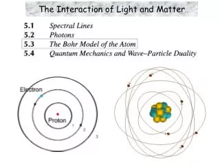
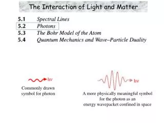
Electronic structures of simple molecule Energy Excited state Singlet S1 Excited state Triplet T1 Vibration states D Dissociated states Bond length S0 Ground state
S1 T1 D S0 S1 transition S0 Interaction between photon and molecule S1 T1 UV-vis F A P IR S0
Key concept from energy diagram Electronic structures Singlet and triplet Bond length for ground and excited states Vibrational structures-infrared absorption/transmission (FTIR) Internal conversion Intersystem crossing Photon adsorption excitation (Beer’s law, UV-vis) Frank Condon condition and The Stokes' shift Radionless relaxation and vibration relaxation Luminescence-fluorescence/phosphorescence
Type of optical spectroscopy UV-vis absorption spectroscopy (UV-Vis) FT-IR absorption/transmission spectroscopy (FTIR) Atomic absorption spectroscopy (AAS) Atomic fluorescence spectroscopy (AFS) X-ray fluorescence spectroscopy (XFS) What you will learn: The excitation mechanism Monochromator design Instrument principle Quantitative methods
Excitation sources Detectors Deuterium Lamp PMT Tungsten Lamp CCD/CID Monochromators Laser Photodiode Filters X-ray tube Grating+slit Thermocouple Mercury lamp prism MCT Xenon lamp Pyroelectric detector Silicon carbide globar Flame Furnaces Plasmas Hollow-cathode lamp Optical spectrophotometer components UV UV-vis X-ray, UV, vis, IR X-ray UV-vis UV-vis IR What is the advantage and disadvantage?
Sources • Continuous Source • Line Source • Pulsed Continuum Line
Mercury Arc Xenon Arc Tungsten
Light sources Brightness Line width Background Stability Lifetime What is the important properties of a source? Black-body radiation for vis and IR but not UV - a tungsten lamp is an excellent source of black-body radiation - operates at 3000 K - produces l from 320 to 2500 nm ( How much in cm-1, J, Hz and eV?) For UV: - a common lamp is a deuterium arc lamp - electric discharge causes D2 to dissociate and emit UV radiation (160 – 325 nm) - other good sources are: Xe (250 – 1000 nm) Hg (280 – 1400 nm) Lasers: - high power - very good for studying reactions - narrow line width - coherence - can fine-tune the desired wavelength (but choice of wavelength is limited) - £££ expensive £££
Design of optical spectrophotometers Single Beam vs. Double Beam (a) single-beam design (b) dual channel design with beams separated in space but simultaneous in time (c) double-beam design in which beams alternate between two channels."
Sample a source containers:for UV: quartz (won’t block out the light) for vis: glass [l 800nm (red) to l 400 nm (violet)] for IR: NaCl (to or 15384 nm or 650 cm-1) KBr (to 22222 nm or 450 cm-1) CsI (to 50000 nm or 200 cm-1) Best material: diamond, why? Optical transmission coefficient Criteria High transmission Chemically inert Mechanically strong
Monochromators Early spectrophotometers used prisms - quartz for UV - glass for vis and IR These are now superseded by: Diffraction gratings: - made by drawing lines on a glass with a diamond stylus ca. 20 grooves mm-1 for far IR ca. 6000 mm-1 for UV/vis - can use plastic replicas in less expensive instruments Think of diffraction on a CD http://www.ii.com/images/prism.jpg http://www.mrfiber.com/images/cddiffract.jpg 10mmx10mm http://www.veeco.com/library/nanotheater_detail.php?type=application&id=331&app_id=34
Monochromators Polychromatic radiation enters The light is collimated the first concave mirror Reflection grating diffracts different wavelengths at different angles Second concave mirror focuses each wavelength at different point of focal plane Orientation of the reflection grating directs only one narrow band of wavelengths to exit slit http://oco.jpl.nasa.gov/images/grating_spec-br.jpg
Interference in diffraction d sin(q)+d sin(f)=nl d Bragg condition Phase relationship q>0 f<0 f q n=1, 2, 3 In-phase n=1/2, 3/2, 5/2 out-phase
Monochromators: reflection grating Each wavelength is diffracted off the grating at a different angle Angle of deviation of diffracted beam is wavelength dependent diffraction grating separates the incident beam into its constituent wavelengths components Groove dimensions and spacings are on the order of the wavelength in question In order for the emerging light to be of any use, the emerging light beams must be in phase with each other l Resolution of grating: n: diffraction order N: number of illuminated groves =nN Dl Angular resolution: As: d sin(q)+d sin(f)=nl So: n Dl=d cos(f) Df Therefore: Df/Dl=n/[d cos(f)]
Monochromators: slit Bottom line: - it is usually possible to arrange slits and mirrors so that the first order (n = 1) reflection is separated - a waveband of ca. 0.2 nm is obtainable However, the slit width determines the resolution and signal to noise ratio Large slit width: more energy reaching the detector higher signal:noise Small slit width: less energy reaching the detector BUT better resolution!
Detectors : Radiation-----charger converter Choice of detector depends upon what wavelength you are studying Want the best response for the wavelength (or wavelength range) that you are studying In a single-beam spectrophotometer, the 100% transmittance control must be adjusted each time the wavelength is changed In a double-beam spectrophotometer, this is done for you!
Photomultiplier-single channel, but very high sensitivity - Light falls on a photosensitive alloy (Cs3Sb, K2CsSb, Na2KSb) - Electrons from surface are accelerated towards secondary electrodes called dynodes and gain enough energy to remove further electrons (typically 4-12, to 50 with GaP). - For 9 stages giving 4 electrons for 1, the amplification is 49 or 2.6 x 10⁵) - The output is fed to an amplifier which generates a signal - To minimise noise it is necessary to operate at the lowest possible voltage
Photodiode Array-multiplex, but low sensitivity Good for quick (fraction of a second) scanning of a full spectrum Uses semiconductor material: Remember: n-type silicon has a conduction electron – P or As doped p-type silicon has a ‘hole’ or electron vacancy – Al or B doped A diode is a pn junction: under forward bias, current flows from n-Si to p-Si under reverse bias, no current flows boundary is called a depletion layer or region
Photodiode Array - Electrons excited by light partially discharge the condenser - Current which is necessary to restore the charge can be detected - The more radiation that strikes, the less charge remains - Less sensitive than photomultipliers several placed on placed on single crystal - Different wavelengths can be directed to different diodes - Good for 500 to 1100 nm - For some crystals (i.e. HgCdTe) the response time is about 50 ns
Photodiode Array Spectrophotometer - For photodiode array spectrophotometers, a white light passes through sample - The grating polychromator disperses the light into the component wavelengths - All wavelengths are measured simultaneously - Resolution depends upon the distance between the diodes and amount of dispersion No moving parts! Simple mechanical and optical design, very compact.
Photodiode Array Spectrophotometers vs Dispersive Spectrophotometers Dispersive Spectrophotometer: - only a narrow band of wavelengths reaches the detector at a time - slow spectral acquisition (ca. 1 min) - several moving parts (gratings, filters, mirrors, etc.) - resolution: ca. 0.1 nm - produces less stray light greater dynamic range for measuring high absorbance - sensitive to stray light from outside sources i.e. room light Photodiode Array Spectrophotometer: - no moving parts rugged - faster spectral acquisition (ca. 1 sec) - not dramatically affect by room light What are the components 1 to 10? From: http://www.oceanoptics.com/