Download
1 / 46
610 likes | 797 Views
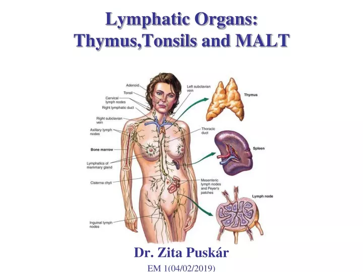
Lymphatic Organs: Thymus,Tonsils and MALT. Dr. Zita Puskár EM 1(04/02/2019). The Immune System. Function: distinguishing between self or non - self , dangerous or non-dangerous and responding to those with tolerance or elimination .

E N D
Lymphatic Organs: Thymus,Tonsils and MALT Dr. Zita Puskár EM 1(04/02/2019)
The Immune System Function: distinguishing between self or non-self, dangerous or non-dangerous and responding to those with tolerance or elimination. Immunohomeostasis - maintenance of the genomic permanency • Defense against the pathogens • Elimination of the transformed self structures Antigen: every structure (cells, molecules, microbes) that the immune system recognizes and responds to it
Innate and Adaptive Immune System Innate Cells → cellular immune response Monocytes - macrophages Granulocytes Dendritic cells Mast cells Soluble molecules → humoral immune response Complement proteins (glycoproteins, enzymes, receptors) Adaptive Cells → cellular immune response B lymphocytes T lymphocytes Soluble molecules → humoral immune response Antibodies
Antigen Processing Phagocytosis, antigen presentation, cell activation – „professional” antigen presenting cells (APCs): macrophage, dendritic cell • Phagocytosis, antigen presentation, cell activation oxygen burst (bacterial infections) – neutrophil granulocyte (microphage) • Elimination of helminthic worms and protozoa, (MBP toxicity, ECP neurotoxicity), allergic reaction with inflammation – eosinophil granulocyte • Allergic reaction, defence against parasites – mast cell, basophil granulocyte • Elimination of viral infected and tumour cells – Natural Killer Cell (NK)
Activation of the Innate System Recognized structures: Pathogen Associated Molecular Pattern (PAMP) Receptors: Pattern Recognizing Receptors (PRR). They are not clonal. They are the same on different cell types. Fc receptors, binding antigen-antibody complexes. PPR (Pattern recognition Receptors) (only examples) Mannose receptor Scavenger receptor CD 14 Toll-like receptors
Major Histocompatibility Complex:MHC-I and -II Function: Determination the immunological „self”, helping the formation of T-cell repertoire and the recognition of proteins by T-cells (membrane glycoproteins – peptidereceptors) Peptides that join the MHC I consist of 8-10 amino acids. Cells that express MHC-I–allnucleated cell Peptides that join the MHC II contain 13-23 amino acid. Cells that express MHC-II –B cell, macrophage, dendritic cell (APC), thymic epithel Human MHC-HLA: Human Leucocyte Antigen complex
Antigen Presentation Antigens originated from lipids join to CD1, peptide antigens join to MHC-I or MHC-II. Endogenous proteins (the host cell synthesizes foreign proteins with tumour cell like characteristics in viral or bacterial infected cells). Antigen processing: proteasome (enzymes) → ER (peptide fragments join to MHC-I→ Golgi → vesicular transport → appearance on the cell surface Exogenous proteins (are taken up by APCs by fagocytosis, pinocytosis or receptor mediated endocytosis) Antigen processing: endo- and lysosome system → protected MHC II - formed in the ER join to vesicular system → protein binds to MHC-II → appearance on the cell surface
Antigen Presentation https://www.youtube.com/watch?v=wYPlc6-nnzU
Activation of NK cells NK cells express „Killer Activating Receptor (KAR) „ and „Killer Inhibitory Receptor” (KIR). Binding „self” MHC-I molecules to the KIRs inhibitsthe KARs. NK cell does not destroy self. Virus infected and tumour cells decrease the MHC-I expression to hide themselves from cytotoxic T-cells. Therefore there is no enough MHC-I to activate the KIR so the NK cells attack.
Nomenclature CD (Clusters of Differentiation) – nomenclature of the cell surface molecules that determine the type of the cell, the stage of the differentiation or activation. Cell line markers (eg. Hemopoietic stem cell CD34+, T-helper cell - CD4+, T-citotoxic-CD8+ • Maturation markers (Tymocytes in the thymus CD1+, mature T lymphocyte does not express this) • Activation markers (molecules appearing after stimulation pl. CD25) Cytokines: small peptides or glycoproteins that regulate the cell functions in the immune system through receptor mediated pathways. Lymphokines-cytokines produced by lymphoid cells.
Antibodies – Immunoglobulins (Ig) monomer IgM: naive B cell antigen binding receptor IgD : naive B cell antigen binding receptor IgG: „switched” B cell antigen binding receptor (opsonisation, complement activation, maternal immunity) IgA: „switched” B cell antigen binding receptor (mucosa immunity) IgE: „switched” B cell antigen binding receptor (immediate hypersensitivity) dimer pentamer trimer
B-cell Receptor (BCR) and B-cell Activation BCR: membrane bound immunoglobulin molecule with signal transduction chains. BCR reacts with soluble or corpuscular antigens. B-lymphocyte activation: T dependent (antigen bindingand interaction with T helper cells and cytokines) or T independent (antigen binding and PRR activation and interaction with cytokines)activation results in division and differentiation of B cells into plasma cells and memory B cells.
T-cell Receptor (TCR) and T-cell Activation TCRs: coded by genes belonging to the Ig super-family. They recognize only linear sequence of peptide fragments that join to MHC molecules!!! Real physical interaction is necessary! TCR-MHC-antigen binding is necessary but not enough for T-cell activation. Co-stimulatory molecules are needed!!!
T-cell Classes Production of cytokines (lymphokines) – activation of macrophages, regulation of inflammatory and cytotoxic processes, defense against intracellular pathogens, cellular immune response MHC-II-exogene antigen Th1 Production of lymphokines – activation of B cells, differentiation into plasma cells, defense against extracellular pathogens, humoral immune response Th2 CD4+ Treg Production of lympholines – regulation of immune response Th: helper T-cell Treg: regulatory T-cell Tcit : cytotoxic T-cell Destroys virus or intracellular pathogen infected or tumour cells (perforin, granular enzymes.) CD8+ Tcyt MHC-I-endogeneous antigen
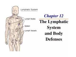
Organization Levels of the Immune System Cells: cells of the innate and adaptive immune system Tissues: blood and lymphatic tissues Organs: lymphatic organs
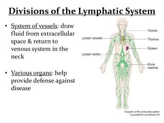
Organization of Lymphatic Organs Primary (central) lymphatic organs Bone marrow: formation of T and B lymphocyte and maturation of B-cells Thymus (thymus gland): maturation ofT-cells Secondary (peripheral) lymphatic organs: „meeting with the antigens” → activation of B and T cells Mucosa Associated Lymphatic Tissue (MALT:antigen in the mucosa) Gastriontestinal tract (digestive tract) GALTTonsils Peyer’s patches in the wall of the intestine Appendix vermiformis Nasal (NALT), Bronchoalveolar (BALT) and urogenital system associated lymphatic tissue Skin Associated lymphatic Tissue (SALT), antigen in the skin) Lymph and lymph node (antigen in tissues) Spleen (antigen in the blood)
Different Forms of Lymphatic Tissue Diffuse lymphoid elements (lymphocytes) Solitary lymph nodule or follicle Epithelium related lymphatic tissue : tonsils Aggregated lymph nodules (pl. Payer’s plaque) Capsulated lymphatic organs spleen Lymph node thymus
Primary Lymphatic Organ: Thymus Arteries: a. thoracica interna, a. pericardiophrenica, a. thyroidea sup. et. inf. • Veins: v. brachiocephalica • Lymph vessels: only efferents!!! Anterior mediastinal lymph nodes • Innervation: n. vagus (parasympatic) and sympatic innervation It is located in the mediastinum supracardiacum (between the sternum and the large vessels)
Histology of the Thymus Lobules Two lobes Capsule : connective tissue (collagen and elastic fibres, plasma and mast cells) blood vessels and efferent lymph vessels) Connective tissue trabeculae (interlobular septum) divide the lobes into lobules. Each lobule has a peripheral darkly stained region called cortex and a central light area called the medulla.
Cells of the Thymus Cytoreticulum: 3D network of epithelial reticular cells with endo- (and ectodermal) origine. These cells are stellate shaped having large euchromatic nuclei and long processes and contain intermedier keratin filaments. They are connected with each other by desmosomes and occluding junctions. Lymphocytes in different developmental stages are among them. 1. capsula 3. trabecula with vessels Subcapsular epithelium Cortex: (5) cortical epithelial reticular cells secreting thymoxatin, thymosin, thymopoietin (2) nurse cells (expressing MHC-II) and T lymphocytes called thymocytes develope among their processes), (9) macrophage Cortico-medullary junction: (6) medullary epithelial reticular cells (larger than the cortical ones, PTH, CRP, inzulin), (7) dendritic cells (APC from the bone marrow), macrophages, eosinophils Medulla: (6) medullary epithelial reticular cells, Hassal- body (keratinized epithelial cells), macrophage, dendritic cells, Hammar myoid cell
Epithelial Reticular Cells, Hassal body Hassal body: concentrically located flattened, eosinophilic, epithelial reticular cells connected with desmosomes. Keratohyalin and cytokeratin are accumulated in the centre. Watanabe et al: Hassall's corpuscles instruct dendritic cells to induce CD4+CD25+ regulatory T cells in human thymus, Naturevolume436, pages1181–1185 (25 August 2005) , Thymic Stromal Lymphopoietin (TSLP)
Dendritic Cells (DC) DC-s originate from the bone marrow. These cells have long processes. The nucleus is irregular, segmented and the cellular organelles surround it.APC! The award of 2011 Nobel Prize for Physiology or Medicine demonstrates the significance of this cell type in health and disease. The 2011 Prize was divided, one half to Ralph M. Steinman "for his discovery of the dendritic cell and its role in adaptive immunity" and the other half jointly to Bruce A. Beutler and Jules A. Hoffmann "for their discoveries concerning the activation of innate immunity." Bruce Beutler was the first to isolate the cytokine tumor necrosis factor (TNF) and later developed the soluble TNF receptor, which is now marketed as Etanercept. Jules Hoffmann identified Toll receptors and their function in innate immune response.
Blood circulation of the Thymus and High Endothelial Venules (HEV) Postcapillary venules have unusual endothelial cells that are high cuboidal and protrude into the lumen of the vessel. The nucleus is large, spherical and lightly stained. The surface glycoproteins and integrins of the apical part of the endothels facilitate fast diapedesis of lymphocytes out of the blood. (15-20 000 cell/second)
T pogenitor (pro-T) cells from the bone marrow enter the Thymus through the venules of the cortico-medullary junction → They move into the subcapsular region and intensively proliferate. They do not express either CD8 or CD4 receptor, double negative T cells (DN) → Environmental signal molecules induce the expression of TCR-s (genetic recombination) and CD4 és CD8, double positive T cells (DP) → They migrate toward the medulla and meet with their own MHC molecules presented by nurse cells, epithelial reticular cells, dendritic cells, macrophages). Those thymocytes that recognize at least one of the MHC-s (MHC restriction) will survive. Others (90%) die by apoptosis. This process is called positive selection. Maturation of T cells Thymocytes that can connect to MHC-I they will be CD4-és CD8+ Tc, the others that bind to MHC-II, they become CD4+és CD8- Th . Then the „self antigens” arepresented. All of those that react to „MHC-self petide complex” will die by apoptosis. This process called negative selection. The matured naive T-cells tolerating self, leave the Thymus through the blood vessels of cortico-medulary junction.
Barriers in the Thymus • Tightly arranged subcapsular epithelial cells (3) form a closed sheath under the capsule, around the trabeculae and vessels to form an inpermeable barrier to macromolecules to protect the thymocytes from contact with foreign antigens. • Layers of blood-thymus barrier: • Epithelial reticular cells (contacted with each other by desmosomes and tight junctions) • Basal lamina of the epithelial reticular cells • Perivascular connective tissue septa • Basal lamina of the endothels • Capillary endothels
Age Related Involution of the Thymus Involution of the Thymus is a process when it decreases in size, weight and activity with advancing age. The Thymus has a critical role in establishing the immune system during childhood. After puberty thymic tissue (especially the cortex) is replaced by adipose tissue, (thymus adiposus) however thymocytes are continually produced into old age. Stress, radiation, steroid treatment result in acute involution. L.P. Hale: Ann.Diag.Path. 2004, 8(1) 50-60
Development of Thymus 1st pharyngeal fold 1st pharyngeal arch 2nd pharyngeal fold 1 Foramen cecum Hyoid arch 2 Thyroid and foramen cecum Aorta dorsalis a sinus Entrance to the cervical sinus 3 Thyroid gland Neural tube 1st pharyngeal arch 4 3rd pharyngeal pouch Cervical sinus 4th haryngeal pouch Anlage of the Thymus 9. Thymus (part of the 3rd pharyngeal pouch) 5-6. week. The thymic epithelium begins as two flask-shaped endodermal diverticula that form from the 3. és 4. pharingeal pouches (endodermal origin) from both sides and extend lateralward and backward into the surrounding mesoderm 7-8. week. The two diverticula join cranially and ventrally from the large vessels comming from the heart and a common capsule is formed by the mesenchyme originating from the neuronal crest. Interlobar mesenchymal septa penetrate into the two lobes. 10. Thymus (part of the 4rd pharyngeal pouch)
(10-15. weeks) 10-11 week. Additional cell types (macrophages, dendritic cells) and T-lymphocytes appear around the thymus and break through the basement membrane and penetrate into the epithelial reticular cells. The cytoreticulum is formed. 15. week: The invasion of the lymphocytes result in the formation of the cortex and medulla. Connective tissue septa divides the lobes into lobules . The interlobular trabeculae penetrates till the cortico-medullary junction. (the lobules appear only in the cortex, the medulla remains uniform) Blood and lymph vessels run in the trabeculae and enter the thymus in the cortico-medullary junction.
General Features of Peripheral Lymphatic Organs Stroma: reticular connective tissue that consists of reticular fibers forming 3D networks and reticular cells. Immune cells and accessory cells are densely packed within the reticular connective tissue (lymphoreticular tissue) Cells: lymphoid cells: B and T lymphocytes, plasma cells, NK cells, accessory cells: macrophages, dendritic cells: follicular dendritic cells (FDC): bind native antigens and help the differentiation of B-cells in the germinal center of the lymphatic nodule (B- dependent area) dendritic cells (interdigitating dendritic cell) present MHC-II joined antigens to the a Th cells in the T- dependent area of lymphatic organs, (eg. Langerhans cells of the skin, interstitial dendritic cells in the connective tissue) Vessels: blood vessels (with high endothelial venules, HEV) lymph capillaries Special formations: lymphatic nodules (follicles)
Primary and Secondary Follicles (Lymphatic Nodules) Cap Reticular cell FDC Light zone Germinal center Primary (d=50-100 µm) Germinal center reaction Dark zone Primary: reticulum cells, resting naive B-lymphocytes (antigen free environment, e.g. intrauterine life) Secondary: cap (like the primer follicle). Germinal centre (centrum germinativum): dark zone - B lymphoblasts (centroblasts), dividing forms light zone-smaller centrocytes , follicular dendritic cells, macrophages s v Secondary (d=200-400 µm)
Germinal Centre - Reaction: B-cell activation centrocyta centroblast tingible body macrophage Antigen stimulated B cells interact with specific T cells at the border between B cell zone and T cell zone and migrate into the centre of the follicle. B cells differentiate into centroblasts (dark zone B cells), divide and undergo somatic hypermutations and generate large number of immunoglobulin mutations in the dark zone. Centroblasts differentiate into centrocytes (light zone B cells) expressing high-affinity antibodies and form the light zone. They are selected and instructed to differentiate into memory B cells or plasma cells.
Diversity of the Antigen Binding Receptors Genetic mechanism that results in diversity and specificity is the somatic recombination of genes that code the antigen binding site of the immunoglobulins and T-cell receptors.
T- and B-dependent Areas lymph node spleen tonsil Naive B-cells, macrophages, follicular dendritic cells are the main cell types in the follicles, while naive T cells, dendritic cells, macrophages are located in the interfollicular areas.
MALT: Tonsils Pharyngeal tonsil Waldeyer ring Tubal tonsil Palatine tonsil Lingual tonsil The tonsils of Waladeyer’s ring outline the plane of the former oropharyngeal membrane
K General Features of Tonsils MALT – Mucosa Associated Lymphatic Tissue : • Lymphoepithelial tissue: the epithelium is infiltrated with lymphatic cells • Lymphoreticular connective tissue with lymphatic nodules. The follicles are B-dependent areas. Among the follicles, T-dependent areas with blood vessels (HEV), and lymph capillaries. crypts (fossula, lacuna): epithelial invaginations (increasing the surface for immune response) K K lymphoepithel K K
Histology of LingualTonsil • Epithelium: stratified squamous non-keratinizing epithelium infiltrated with lymphoid cells. • Crypts are not too deep • Lymphoreticular connective tissue with follicles • Mucous lingual glands that release their saliva into the crypts • Skeletal muscle • Adipose tissue
Palatine Tonsil Palatopharingeal arch tonsillar fossa Palatoglossal arch
Histology of Palatine Tonsil hemicapsule • Epithelium: stratified squamous non-keratinizing epithelium infiltrated with lymphoid cells. • Crypts are deep and branched (during bacterial infection (strepto- or staphylococcus) the crypts are filled with neutrophils → pus • Lymphoreticular connective tissue with follicles • Hemicapsule: a band of dense connective tissue acting as a capsule separating the lymphoid tissue from the subjacent structure. Function: barrier against to spread infections (area for tonsillectomy)
Tonsillitis, Tonsillectomy physiological inflammed tonsillectomy follicular tonsillitis
Pharyngeal Tonsil In the pharyngeal fornix
Histology of Pharyngeal Tonsil Structure: 6-8 sagittal folds and deep pits among the folds Epithelium: ciliated pseudostratified columnar epithelium (with islands of stratified squamous non-keratinizing epithelium on the top of the folds and in the pits) infiltrated with lymphoid cells. The stuctural composition of the tubal tonsil is very similar to the pharyngeal tonsil but cartilage is located under the follicle filled connective tissue in this case.
MALT: Peyer’s Patches ileum
References: Anthony L. Mescher: Junqueira’s Basic Histology, New York, 2010 Michael Ross and Lynn J. Romrell: Histology, Baltimore, 1989 Buzás Edit, Falus András, Rajnavölgyi Éva: Az immunológia alapjai, Budapest, 2007 Röhlich Pál: Szövettan. Budapest, 2006 Erdei Anna, Sármay Gabriella, Prechl József: Immunológia , Budapest, 2012