Download
1 / 2
20 likes | 202 Views
Quantifying Local Shears in Pure Ti grains with SEM and Crystal Plasticity Finite Element (CPFE) Simulation to Assess Damage Thomas R. Bieler, Michigan State University, DMR 1108211. Prism slip. Basal slip.

E N D
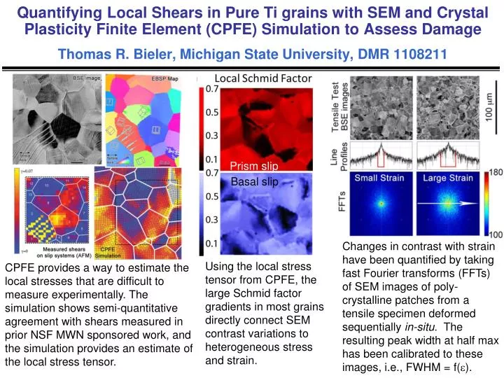
Quantifying Local Shears in Pure Ti grains with SEM and Crystal Plasticity Finite Element (CPFE) Simulation to Assess DamageThomas R. Bieler, Michigan State University, DMR 1108211 Prism slip Basal slip Changes in contrast with strain have been quantified by taking fast Fourier transforms (FFTs) of SEM images of poly-crystalline patches from a tensile specimen deformed sequentially in-situ. The resulting peak width at half max has been calibrated to these images, i.e., FWHM = f(). Using the localstress tensor from CPFE, the large Schmid factor gradients in most grainsdirectly connect SEM contrast variations to heterogeneous stress and strain. CPFE provides a way to estimate the local stresses that are difficult to measure experimentally. The simulation shows semi-quantitative agreement with shears measured in prior NSF MWN sponsored work, and the simulation provides an estimateof the local stress tensor.
Quantifying Local Shear in Pure Ti grains with SEM and Crystal Plasticity Finite Element (CPFE) Simulation to Assess DamageThomas R. Bieler, Michigan State University, DMR 1108211 Fatigue Crack tip Before Fatigue 30 mm BSE images show changes in channeling contrast with strain Evolution of mesoscale plastic strain ahead of a fatigue crack in pure Ti was quantified using the FFTs of backscattered electron contrast in a 15x15 array of images collected in the crack tip region. This characterization of the strain field was compared to the zone of plastic work measured using thermal-elastic stress analysis (TSA) using a sensitive thermal camera. Comparison with analytical solutions shows no agreement with the Von Mises analysis, but good agreement with Dugdaleand Irwinanalyses. These results indicate that both TSA and the approach developed here are reasonably robust and can be applied to a broader range of loading geometries, providing a diagnostic measure of damage that can impact the design of complex industrial loading and forming operations where accurate measurements of strain fields and damage is required.