Download
1 / 60
1.49k likes | 4.08k Views
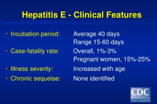
C.S.O.M.: Clinical Features. Dr. Vishal Sharma. Definition. Chronic (> 3 months) pyogenic infection of middle ear cleft mucosa , characterized by persistent perforation of tympanic membrane, ear discharge & decreased hearing Prevalence in Nepal: 7.2 %. Types of C.S.O.M.

E N D
C.S.O.M.: Clinical Features Dr. Vishal Sharma
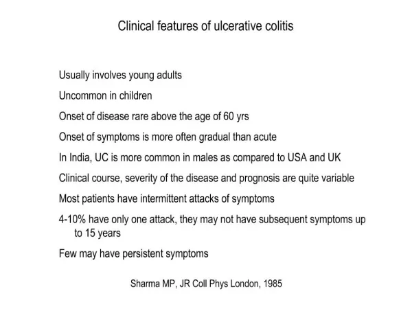
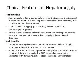
Definition • Chronic (> 3 months) pyogenic infection of middle ear cleft mucosa, characterized by persistent perforation of tympanic membrane, ear discharge & decreased hearing • Prevalence in Nepal: 7.2 %
Types of C.S.O.M. Tubo-tympanic: chronic pyogenic infection of middle ear cleft mucosa with persistent perforation in pars tensa Attico-antral:chronic pyogenic infection of middle ear cleft with cholesteatoma & granulations in attic or postero-superior quadrant of pars tensa
Types Perforation of Pars Tensa 1. Central tubo-tympanic Small Medium Large Subtotal 2. Central with ingrowing epithelium attico-antral 3. Marginal attico-antral 4. Total attico-antral Perforation of Pars Flaccida 1. Attic attico-antral
4 quadrants of T.M. umbo
Small perforation Involves only one quadrant or < 10% of pars tensa
Medium perforation Involves two quadrants or 10 – 40 % of pars tensa
Large perforation Involves 3 or 4 quadrants with wide T.M. remnant or > 40 % of pars tensa
Subtotal perforation Involves all 4 quadrants & reaches up to annulus fibrosus
In growing epithelium T.M. perforation with inward migration of epithelium
Marginal perforation Erodes annulus fibrosus & one margin is formed by bony tympanic annulus
Total perforation Total erosion of pars tensa & anulus fibrosus
Attic perforation Involves pars flaccida
Grade 1 retraction • Dull, lustreless T.M. • Prominent annulus • Cone of light absent • Handle medialized • Prominent lateral process • Malleolar folds sickle shaped
Grade 2 retraction Eardrum touches incus
Grade 3 retraction TM touches promontory (atelectasis) but mobile on Valsalva maneuver or Siegalization
Grade 4 retraction TM firmly adherent to promontory & immobile on Valsalva maneuver or Siegalization
Otological examination 1. Pre-auricular region: sinus, lymph node 2. Pinna: size, position, deformity, swelling 3. Post-auricular region: surgical scar, swelling, fistula, lymph node 4. External auditory canal: meatal opening, otitis externa, wax, fungal debris, ear discharge
Otological examination 5. Tympanic membrane: intact:colour, position, mobility, tympanosclerosis, retraction pocket perforated:type, site, size & margin of perforationhandle of malleus; middle ear cavity (mucosa, ear discharge, polyp, granulations, cholesteatoma flakes); pars flaccida
Otological examination 6. Mastoid cavity: size, facial ridge, discharge, epithelialization, granulations, polyps 7. Tragal tenderness: associated otitis externa 8. Mastoid tenderness: cymba conchae, mastoid body + tip & posterior zygoma root 9. Fistula sign 10. Facial nerve function 11. Tuning Fork Tests
Predisposing factors • Upper respiratory tract infection (recurrent) • Upper respiratory tract allergy • Pre-existing otitis media with effusion • Cleft palate • Immune deficiency: diabetes, AIDS • Poor socio-economic status
Bacteria responsible • Staphylococcus aureus • Pseudomonas aeruginosa • Klebsiella • Proteus • Streptococcus • Bacteroides
Routes of infection • Via Eustachian tube: U.R.T.I., nose blowing, regurgitation of milk • Via tympanic membrane perforation:following A.S.O.M. or post-traumatic • Haematogenous (rare): viral exanthematous fevers
Pathological Changes 1. Eardrum: central perforation; myringosclerosis 2. Ossicles:Destruction (hyperaemic decalcification) Tympanoslerosis Fibrosis + Adhesions 3. Middle ear mucosa:edematous, pale pink 4. Mastoid bone:sclerosis
Clinical Features Ear discharge:profuse, mucoid / muco-purulent, intermittent, odourless, not blood-stained Hearing Loss: usually conductive (25-50 dB) absent in small, dry perforations round window shielding by ear discharge leads to better hearing Tympanic membrane:central perforation
Cholesteatoma • Term used by Johannes Müller in 1858 • Three dimensional sac lined by matrix of keratinizing stratified squamous epithelium which rests on a thin layer of fibrous tissue • Contains desquamated keratin debris • Grows at the expense of surrounding bone • Not a tumor & has no cholesterol • Epidermosis is a better term
Causes of bone destruction 1. Hyperaemic decalcification 2. Osteoclastic bone resorption due to: Acid phosphatase Collagenase Acid proteases Proteolytic enzymes Leukotrienes Cytokines 3. Pressure necrosis: No role 4. Bacterial toxins: No role
Types of Cholesteatoma Congenital (McKenzie) Primary AcquiredSecondary Acquired 1. Retraction pocket 1. Squamous metaplasia (Wittmaack) 2. Epithelial migration 2. Basal cell hyperplasia (Habermann) (Ruedi) Tertiary Acquired 3. Squamous metaplasia 1. Post-traumatic (Sade) 2. Post-tympanoplasty
Congenital cholesteatoma Persistence of congenital cell rests in middle ear, petrous apex, cerebello-pontine angle
Retraction pocket formation Retraction pocket in pars flaccida or Postero-superior quadrant pars tensa due to E.T. dysfunction
Basal cell hyperplasia Hyperplasia of basal cells in epithelial layer of T.M. & their invasion of sub-epithelial tissues
Primary squamous metaplasia Transformation of middle ear mucosa into squamous epithelium due to infection, with no T.M. perforation
Secondary squamous metaplasia Transformation of middle ear mucosa into squamous epithelium due to infection via T.M. perforation
Epithelial migration Migration of epithelium via T.M. perforation into middle ear
Post-traumatic cholesteatoma Mechanisms: 1. Epithelial entrapment in fracture line 2. In growth of epithelium through fracture line 3. Traumatic implantation of epithelium into middle ear 4. Trapping of epithelium medial to E.A.C. stenosis
Pathological Changes 1. T.M. perforation:marginal or attic 2. T.M. retraction pocket:attic or P.S.Q. 3. Cholesteatoma formation 4. Ossicles:destruction 5. Middle ear mucosa:edematous, red 6. Aural polyp:red, fleshy 7. Osteitis & granulation tissue formation 8. Mastoid bone:erosion, sclerosis