Download
1 / 48
500 likes | 947 Views
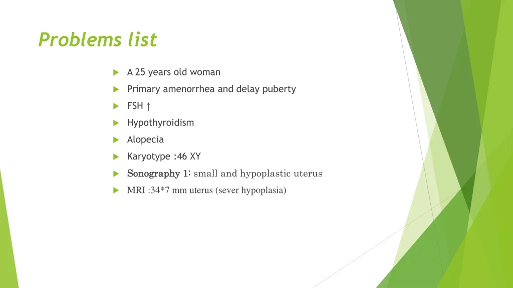
Problems list. A 25 years old woman Primary amenorrhea and delay puberty FSH ↑ Hypothyroidism Alopecia Karyotype :46 XY Sonography 1: small and hypoplastic uterus MRI :34*7 mm uterus (sever hypoplasia ).

E N D
Problems list • A 25 years old woman • Primary amenorrhea and delay puberty • FSH ↑ • Hypothyroidism • Alopecia • Karyotype :46 XY • Sonography 1: small and hypoplastic uterus • MRI :34*7 mm uterus (sever hypoplasia)
A series of sequential diagnostic steps help with the precise identification of a cause of primary amenorrhea • Step 1: History • Has she completed other stages of puberty, including a growth spurt, development of axillary and pubic hair, apocrine sweat glands, and breast development? Lack of pubertal development suggests ovarian or pituitary failure or a chromosomal abnormality. • Is there a family history of delayed or absent puberty (suggesting a possible familial disorder)? • What is the woman's height relative to family members? Short stature may indicate Turner syndrome or hypothalamic-pituitary disease. • Was neonatal and childhood health normal? Neonatal crisis suggests congenital adrenal hyperplasia. • Are there any symptoms of virilization? The presence of virilization suggests polycystic ovary syndrome, an androgen-secreting ovarian or adrenal tumor, or the presence of Y chromosome material. • Lately, has there been stress, change in weight, diet, or exercise habits, or illness that might result in hypothalamic amenorrhea? • Is she taking any drugs that might cause or be associated with amenorrhea? • Is there galactorrhea (suggestive of excess prolactin)? • Are there symptoms of other hypothalamic-pituitary disease, including headaches, visual field defects, fatigue, or polyuria and polydipsia?
Step 2: Physical examination • An evaluation of pubertal development, including current height, weight, and arm span (normal arm span for adults is within 5 cm of height) • An assessment of breast development (eg, by Tanner staging) • A careful genital examination should be performed for clitoral size, pubertal hair development, intactness of the hymen, depth of the vagina, and presence of a cervix, uterus, and ovaries. • Examination of the skin for hirsutism, acne, striae, increased pigmentation, and vitiligo. • Evaluation for the classic physical features of Turner syndrome such as low hair line, web neck, shield chest, and widely spaced nipples should be noted.
Step 3: Basic laboratory testing • This includes measurement of serum beta human chorionic gonadotropin to exclude pregnancy and of serum FSH, and perhaps other hormones. • A high serum FSH concentration is indicative of primary ovarian failure. A karyotype is then required and may demonstrate complete or partial deletion of the X chromosome (Turner syndrome) or the presence of Y chromatin.
Step 4: Imaging • Search for mullerian or wollfian duct by sonograghy and MRI
46 XY disorders of sex development (DSD) Omid GharoeiAhangar
definition • The term disorders of sex development (DSD) includes congenital conditions in which development of chromosomal, gonadal or anatomical sex is atypical.
The 46,XY DSD are characterized by atypical or female external genitalia, caused by incomplete intrauterine masculinization with or without the presence of Müllerian structures. • Male gonads are identified in the majority of 46,XY DSD patients, but in some of them no gonadal tissue is found. • Complete absence of virilization results in normal female external genitalia and these patients generally seek medical attention at pubertal age, due to the absence of breast development and/or primary amenorrhea.
Investigation of DSD patients • Ultrasonography is always the first and often the most valuable imaging modality in DSD patients’ investigation. Ultrasound shows the presence or absence of Müllerian structures at all ages and can locate the gonads and characterize their echo texture. This exam can also identify associated malformations such as kidney abnormalities.
Cytogenetic and Molecular Investigation • The genetic evaluation includes karyotype, FISH and specific molecular studies to screen for the presence of mutations or gene dosage imbalance.
Gonadal Agenesis • Total absence of gonadal tissue confirmed by laparoscopy has rarely been described in XY subjects with female external and internal genitalia indicating the absence of testicular determination. • Mendoncaet al described a pair of siblings, one XY and the other XX, born to a consanguineous marriage, with normal female external and internal genitalia associated to gonadal agenesis. The origin of this disorder remains to be determined.
Complete and partial 46,XY gonadal dysgenesis • The complete form of gonadal dysgenesis was first described by Swyer et al. and is characterized by female external and internal genitalia, lack of secondary sexual characteristics, normal or tall stature without somatic stigmata of Turner syndrome, eunuchoid habitus and the presence of bilateral dysgenetic gonads in XY subjects. Mild clitoromegaly is present in some cases. • The partial form of this syndrome is characterized by variable degrees of impaired testicular development and of testicular function. These patients present a spectrum of atypical genitalia with or without Müllerian structures. Similar phenotypes can also result from a 45,X/46,XY karyotype.
Serum gonadotropin levels are elevated in both the complete and partial forms, mainly FSH levels, which predominate over LH serum levels. Testosterone levels are at prepubertal range in the complete form. Meanwhile, in the partial form, it can range from prepubertal levels to normal adult male levels. • Regarding the genetic etiology, 46,XY gonadal dysgenesis is heterogeneous and can results from defects of any gene involved in the process of gonadal formation. Mutations inNR5A1, MAP3K1 and SRYare the most frequent molecular cause of 46,XY gonadal dysgenesis.
Dysgenetic 46,XY DSD due to MAP3K1 underexpression • MAPK signaling pathway role in mammalian sex-determination is still poorly understood. • In mice, it has been shown that the Map3k4 gene is essential for testicular determination, since the lack of activity of this protein leads to failure of testicular cord development and disorganization of gonadal tissue in formation. • In mice, the reduction of the Gadd45/Map3k4/p38 pathway activity is associated with a reduction in the Sry expression in the XY mice gonad at sex-determination causing sex-reversal in these animals.
46,XY DSD due to the underexpression of steroidogenic factor-1 (NR5A1/SF1) • The reviewed data of seventy-two 46,XY DSD patients with NR5A1 mutations reported in the literature, suggested that Müllerian derivatives are present in about 24% of the cases. However, persistently elevated FSH levels after puberty found in all patients studied suggest an impairment of Sertoli cells function in post pubertal age. • NR5A1 defects can be found in association with a wide range of human reproductive phenotypes such as 46,XY and 46,XX disorders of sex development (DSD) associated or not with primary adrenal insufficiency, male infertility, primary ovarian insufficiency.and finally testicular or ovariotesticular 46,XX DSD.
46,XYDSD due to underexpression of SRYgene Most of the authors reported mutations in SRY gene in less than 20% of the patients with complete 46,XY gonadal dysgenesis. are predominantly de novo mutations. However, some cases of fertile fathers and their XY affected children, sharing the same altered SRY sequence, have been reported.
46,XY DSD due to testosterone production defects • A review of the literature allowed to delineate the characteristics of 46,XY DSD due to the complete form of Leydig cell hypoplasia as: • 1) female external genitalia leading to female sex assignment • 2) no development of sexual characteristics at puberty, • 3) undescended testes slightly smaller than normal with relatively preserved seminiferous tubules and absence of mature Leydigcells • 4) presence of rudimentary epididymis and vas deferens and absence of uterus and fallopian tubes • 5) low testosterone levels despite elevated gonadotropin levels, with elevated LH levels predominant over FSH levels • 6) testicular unresponsiveness to hCGstimulation • 7) no abnormal step up in testosterone biosynthesis precursors
46,XY DSD DUE TO ENZYMATIC DEFECTS IN TESTOSTERONE SYNTHESIS • Six enzymatic defects that alter the normal synthesis of testosterone have been described to date. • Three of them are associated with defects in cortisol synthesis leading to congenital adrenal hyperplasia. • All of them present an autosomal recessive mode of inheritance and genetic counseling is mandatory, since the chance of recurring synthesis defects among siblings is 25%.
46,XY DSD DUE TO DEFECTS IN ANDROGEN ACTION • Androgen insensitivity syndrome (AIS) is the most frequent cause of atypical genitalia in individuals with 46,XY karyotype. • The undervirilization can be complete (female external genitalia) or incomplete virilization with a spectrum of atypical genitalia. • Differing degrees of resistance lead to 3 three phenotypes: • a complete form with female normal-appearing external genitalia • a partial form with a wide range of virilization of the external genitalia • a mild form with only oligospermia, infertility and/or micropenis
46,XY DSD DUE TO PERSISTENT MÜLLERIAN DUCT • Defect in AMH Synthesis in AMH Receptor: • Affected patients present a male phenotype, usually along with bilateral cryptorchidism and inguinal hernia. Leydig cell function is preserved, but azoospermia is common due to the malformation of ductus deferens or agenesis of epididymis. When the hernia is surgically corrected, the presence of a uterus, fallopian tubes and superior part of the vagina can be verified.
46,XY ovotesticularDSD • There are rare descriptions of 46,XY DSD patients with well characterized ovarian tissue with primordial follicles and testicular tissue. • To our knowledge there are no descriptions of an adult patient with 46,XY ovotesticular DSD with functioning ovarian tissue, as occurs in all 46,XX ovotesticularDSD. • Therefore the diagnosis of 46,XY ovotesticular DSD is debatable.
Gonadal Tumor Development in 46,XY DSD Patients • Neoplastic transformation of germ cells in dysgenetic gonads (gonadoblastomas and/or an invasive germ cell tumor) occurs in 20-30% of 46,XY DSD patients and is associated with the presence of Y chromosome or part of. • Bilateral gonadectomy should be performed in 46,XY patients before pubertal age to avoid degeneration of dysgenetic tissue, unless the gonad is functional and easily accessible to palpation and imaging studies, which should be performed yearly.
Management of patients with 46,XY DSD • Hormonal Therapy: • The purpose of the hormonal therapy is the development of female sexual characteristics and menses in the patients with uterus. The treatment must simulate normal puberty, by introducing low doses of estrogen at 9-11 years to avoid excessive bone maturation in short children. • Estrogen therapy should be initiated at a low dose (one sixth to one quarter of the adult dose) and increased gradually at intervals of 6 months. The initial dosage of conjugate estrogens (0.07 to 0.15 mg/day orally) or oral or topic 17β-estradiol (0.5 mg daily) is kept as the patient presents progressive breast development. • After breast development is complete, the estrogen dose is maintained at (0.625 mg/day of conjugate estrogen) or 1 mg twice a day of oral or topic 17b-estradiol) continuously and medroxyprogesterone acetate (5 to 10 mg/day) or micronized progesterone 50 mg/day, from the 1st to the 12th day of the month), is added to induce menses.
surgical treatment • The aim of surgical treatment is to allow development of adequate external genitalia and remove internal structures that are inappropriate for the social sex. Patients must undergo surgical treatment preferably before 2 years of age, which is the time when the child becomes aware of his/her genitals and social sex. Only skilled surgeons with specific training in the surgery of DSD should perform these procedures. • In these patients, the indications for laparoscopy are the removal of normal gonads and ductal structures that are contrary to the assigned gender and the removal of dysgenetic gonads, which are nonfunctional and present potential for malignancy.
CLINICAL PRESENTATIONS • The clinical manifestations of testicular regression can be associated with the embryological sequence and can be classified into early and late embryonic, and early, mid and late fetal or early neonatal events. • TRS patients usually exhibit a male phenotype. However, although uncommon, the phenotypic spectrum associated with TRS may vary from normal male with unilateral nonpalpable testis through phenotypic male with micropenis, to phenotypic female. • These genetically male individuals (46, XY) present with unilateral or bilateral absence of detectable testis structures and Mullerian duct system. • Embryonic TRS has been considered to be part of the clinical spectrum of partial 46, XY gonadal dysgenesis.