Download
1 / 17
170 likes | 288 Views
A Model to Determine Molecular Weights of Proteins from Gel Electrophoresis. By Jose Ceja Kamyar Ghods CSUN/JPL-PAIR 2001. Outline. Getting the data (Standards) Choosing a Model Getting the data (Unknowns) Applying the model Results and conclusions. Getting the Data.

E N D
A Model to Determine Molecular Weights of Proteins from Gel Electrophoresis By Jose Ceja Kamyar Ghods CSUN/JPL-PAIR 2001
Outline • Getting the data (Standards) • Choosing a Model • Getting the data (Unknowns) • Applying the model • Results and conclusions
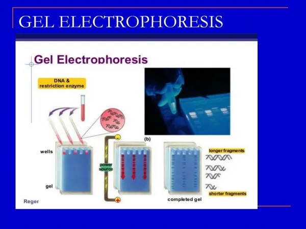
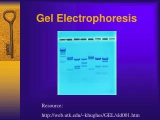
Getting the Data • Two Methods were used: • Adobe Photoshop • Spot Viewer
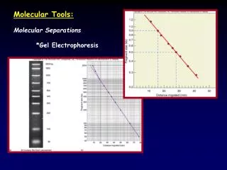
Choosing a Model • Cubic of form: log(MW)=a+b(RM)^2+c(RM)^3 • Cubic of form: log(MW)=a+b(log(RM))^2+c(log(RM))^3 • Quad Cross Validation of form: log(MW)=a+b(RM)+c(RM)^2 • SLIC
Applying Our Model • Collected unknown data using Photoshop • Spot viewer not designed for 1D gels and not well understood. • Applied best cubic model to • each gel.
Applying Our Model • Created an average of our two data sets • Applied cubic model to all • Each standard had 3 cubic fits • Used data that had the best cubic fit for each standard
Jose’s Unknown • Frog skin Gels @ 7 and 12% for males and females • Within the same gel different lanes had different bands. • Male and Female frog’s skin do not have the exact same proteins
Komy’s Unknown • Comparing 3 methods • Overall the Manual method found the most proteins and the Amylase method found the least. • The replicates of each gel were pickkin up more and different proteins.
Conclusions & Future Work • We both found that the higher concentrations found more proteins. • Photoshop is more reliable for dense 1D gels. • Out of the four models we tried, the cubic model was the best one. • Further study is needed to find a true function relating RM to MW.
Aknowledgements • We thank CSUN/JPL-PAIR program, especially Dr. Carrol, Dr. Clevenson, Dr. Shubin, V. Hutchins and J. Handy. • And our fellow students