Download
1 / 41
490 likes | 981 Views
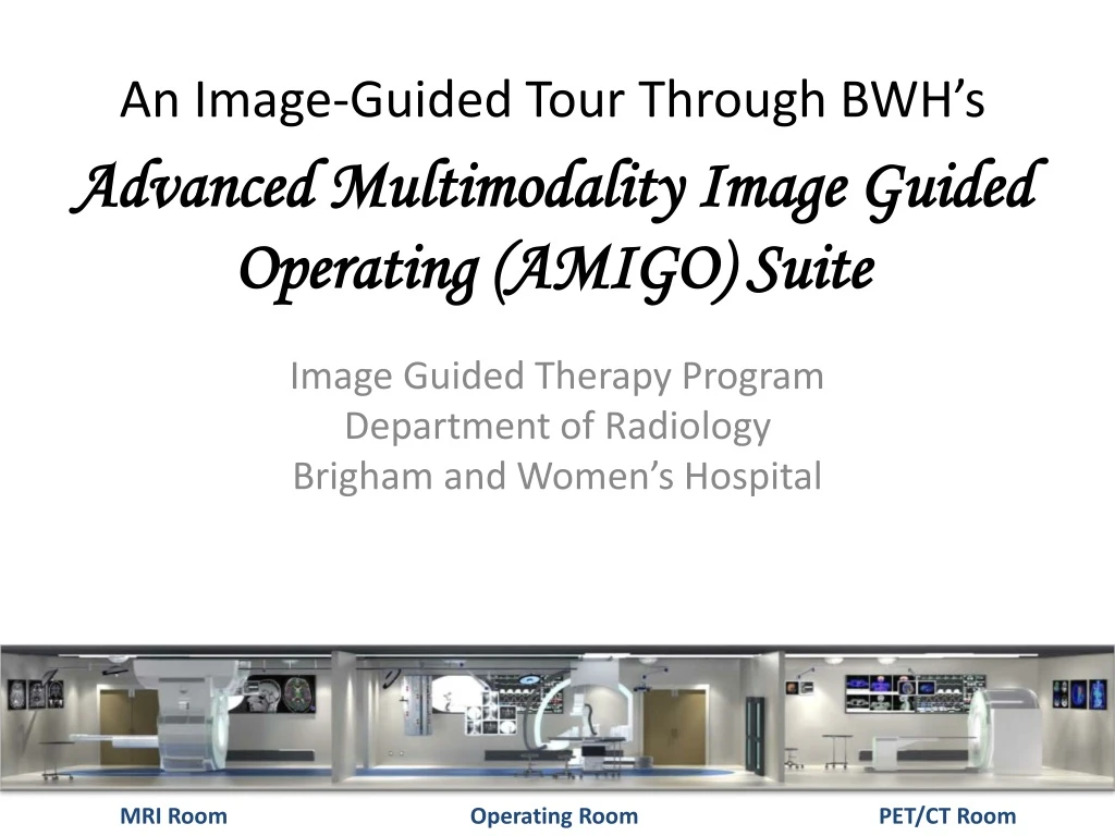
An Image-Guided Tour Through BWH’s Advanced Multimodality Image Guided Operating (AMIGO) Suite. Image Guided Therapy Program Department of Radiology Brigham and Women’s Hospital. MRI Room. Operating Room. PET/CT Room. Supported by.

E N D
An Image-Guided Tour Through BWH’sAdvanced Multimodality Image Guided Operating (AMIGO) Suite Image Guided Therapy Program Department of Radiology Brigham and Women’s Hospital MRI Room Operating Room PET/CT Room
Supported by National Center for Image Guided Therapy (NCIGT) P41 RR019703 (Jolesz, Tempany) 2005-2015
Credits • This presentation researched and created by Mallika Winsor • Photographs by Junichi Tokuda and Dan Kacher
From Conceptualized Sketch… ...To Completed Suite • Advanced Multimodality Image Guided Operating (AMIGO) Suite • P41 RR019703 – National Center for Image Guided Therapy (NCIGT) 2005-2015 • Ferenc Jolesz, MD Clare Tempany, MD
The OR and MR rooms of AMIGO • Advanced Multimodality Image Guided Operating (AMIGO) Suite • P41 RR019703 – National Center for Image Guided Therapy (NCIGT) 2005-2015 • Ferenc Jolesz, MD Clare Tempany, MD
Imaging Equipment in AMIGO • Room 1: MRI Room • Siemens 3T Verio MR scanner that moves along a ceiling track between the MRI room and the OR • Room 2: PET-CT Room • Siemens PET-CT • Room 3: Operating Room • BK Medical Pro Focus UltraView Surgical Ultrasound with Prostate Transducer • Siemens S2000 Ultrasound • Siemens Artis Zee ceiling mounted X-ray Fluoroscopy system with Navigation Package and DynaCT • Zeiss Pentero surgical microscope • Navigation • BrainLAB navigation system • Sentinelle Medical (Hologic) Aegis Navigation Workstation • St Jude Medical mapping and navigation system • IntraMedical Imaging Node Seeker and Beta Probe • Robin Medical Endoscout
The OR and MR rooms of AMIGO • Advanced Multimodality Image Guided Operating (AMIGO) Suite • P41 RR019703 – National Center for Image Guided Therapy (NCIGT) 2005-2015 • Ferenc Jolesz, MD Clare Tempany, MD
Magnetic Resonance Imaging (MRI) Goal: Highly detailed anatomical images What Does It Do? Applications Used to distinguish pathological tissue from normal tissue Comparable resolution, much better contrast resolution (between arbitrarily different but not identical tissues) than CT Advantages Harmless to patient What it uses Strong magnetic fields Non-ionizing radiation • Strong magnetic field is applied, causing the nuclei within the patient’s body to rotate and align • Scanner detects, records, and combines the different rotating fields into a single image • Gradients in all directions are combined to create a 3D image • Strong contrast between different soft tissues within body • Better at producing images of brain, muscles, heart, cancers than CT or x-rays
Magnetic Resonance Imaging (MRI)Terminology Parameters of Image Acquisition Edema Abnormal accumulation of fluid beneath the skin or in cavities of the body Caused by increased secretion of fluid into the interstitium or impaired removal of fluid • TE = echo time • TR = repetition time • TI = inversion time • Time between inversion and excitation pulses
Magnetic Resonance Imaging (MRI)Basic Scans T1-weighted MRI scan T2-weighted MRI scan Highlights fluids Fat = dark; water = light Good for imaging edema Heavily used for clinical scans of cerebral spinal fluid Long TE; long TR • Highlights fat deposits • Water = dark; fat = bright • Good grey v. white matter contrast • Commonly run clinical scan • Short TE; short TR T2*-weighted MRI scan Spin density weighted MRI scan • Gradient Echo (GRE) sequence • Additional loss over T2 decay • Air/tissue boundaries susceptible • Increases contrast of specific tissues • Long TE; long TR • No contrast from either T1 or T2 decay • Uses spin echo (sometimes gradient echo) sequences • Short TE; long TR
Magnetic Resonance Imaging (MRI)Specialized Scans Diffusion MRI scan Fluid Attenuated Inversion Recovery (FLAIR) Inversion-recovery pulse sequence that nulls fluid signals Free water = dark; edematous tissue = bright Most sensitive way to evaluate brain for demyelinating diseases Carefully-selected TI allows signal from any particular tissue to be suppressed Conversion of T2-weighted sequence via additional radio frequency pulse and manipulation of magnetic gradients • Diffusion tensor imaging (DTI) enables diffusion to be measured in multiple directions • Make brain maps of fiber directions • Examine connectivity of different regions of brain • Examine areas of neural degeneration and demyelination Functional MRI (fMRI) • Measures signal changes in the brain due to changing neural activity • Low resolution, rapid rate scans • BOLD (Blood-Oxygen-Level Dependent) effect • Increases in neural activity cause changes in MR signals via T2* changes • Increased neural activity increases demand for oxygen • Vascular system overcompensated, increasing oxygenated hemoglobin • High resolution 3D maps of venous vasculature within neural tissue Real-Time MRI • Continuous monitoring ("filming") of moving objects in real time • Should help provide information about diseases of the joints and heart • May make MRI examinations easier and more comfortable for patients
Magnetic Resonance Imaging (MRI)Medical Applications Interventional Therapy Radiation Therapy Simulation MRIs used to specifically locate tumors within body in preparation for radiation therapy treatments Patient placed in specific, reproducible, body position and scanned MRI system computes precise location, shape and orientation of the tumor mass, correcting for spatial distortion inherent in the system Patient is marked/tattooed with points that, when viewed in specified, reproducible body position, permit precise triangulation for radiation therapy • MRI images used to guide minimally invasive procedures both intraoperatively and interactively • No ferromagnetic instruments used • Some specialized systems allow imaging concurrent with surgery • Usually, surgical procedure is temporarily interrupted so MR images can be acquired to verify success/guide subsequent surgical work
Magnetic Resonance Imaging (MRI) What is MRI Compatibility? Safety Concerns Radio frequency energy Powerful radio transmitter can heat the body to the point of risk of hyperthermia in patients Peripheral Nerve Stimulation Rapid switching on and off of the magnetic field gradients may cause nerve stimulation Acoustic noise Appropriate ear protection is necessary for anyone inside MRI scanner room during examination Cryogens Cryogenic (extremely cold) liquids enable superconducting capabilities of the electromagnetic coils MRI support rooms should be equipped with pressure relief mechanisms and an exhaust fan in case of a quench (shut-down of superconduction electromagnet) Pregnancy Increasingly important for diagnosing and monitoring congenital defects of the fetus (no contrast agents used) Fetal tumors (primarily teratomas) Facilitating open fetal surgery Other fetal interventions Planning to safely deliver and treat babies whose defects would otherwise be fatal • MR-Safe = device or implant that is completely non-magnetic, non-electrically conductive, and non-RF reactive • Titanium and its alloys • MR-Conditional = device that is safe, provided the safety conditions are defined and observed • MR-Unsafe = objects that are significantly ferromagnetic and pose a clear and direct threat to persons and equipment within the magnet room • Patients always asked for complete information about all implants prior to entering the MRI suite
AMIGO’s MRI:Siemens MAGNETOM Verio 3T Who Can It Accommodate? How Does It Help BWH? Minimizes scan rejections due to claustrophobia Reduces necessary sedations Capture sharper images (due to reduced anxiety-related movement) Early access in interventional MRI Opportunities to perform more kinematic studies • Patients up to 550 lbs • Pediatric and elderly patients • Claustrophobic patients • Intensive Care Unit patients • Patients dependent upon medical equipment
AMIGO’s MRI:Siemens MAGNETOM Verio 3T Features → Why this machine? Higher speed, increased image quality Safer scanning environment Increase flexibility, accuracy and speed Enhanced image quality for a wide range of applications Ensures uniform RF distribution in all body regions (optimizes homogeneity) • VQ engine • Ultra-light (6.3 tons) magnet with zero helium boil-off • Total Imaging Matrix (TMI) • TrueForm design • Larger imaging volume, higher image quality, better fat saturation • Whole body imaging up to 6' 4“ • Shortest 3T system available • 70 cm Open Bore • Moves along ceiling into OR Bottom Line: Exceptional diagnostic capabilities, patient comfort, and efficient workflow
A view of OR table and the MR through the PET/CT Bore • Advanced Multimodality Image Guided Operating (AMIGO) Suite • P41 RR019703 – National Center for Image Guided Therapy (NCIGT) 2005-2015 • Ferenc Jolesz, MD Clare Tempany, MD
Positron Emission Tomography-Computed Tomography (PET-CT) Goal: Precise localization of metabolic functions What Does It Do? How Does It Work? Patient fasts for 4 hours or more Intravenous bolus (tracer) injected patient's arm 1-2 hours later, patient placed into PET-CT, usually with arms at the sides or above the head Automatic bed moves head first into the gantry, first obtaining a topogram (whole body flat sagittal section) Operator uses PET-CT console to identify patient and examination, selects the scanning parameters and starts image acquisition period Patient automatically moved head-first into CT gantry, where x-ray tomogram is acquired Patient automatically moved through PET gantry, parallel to CT gantry; PET slices acquired Software reconstructs/aligs PET and CT images Whole body scan (mid-thigh to top of head) takes 5-40 min • Hybrid modality of PET and x-ray CT • Takes both types of images sequentially, in the same session • Images can be combined into a single 2D or 3D superposed image • Detecting changes in molecular activity: • Reveal primary tumors • Detect metastases • Quantify uptake • Improves accuracy in oncology, surgical planning, radiation therapy, cancer staging • Increased image quality, speed, and accuracy of diagnosis
Positron Emission Tomography-Computed Tomography (PET-CT) Positron Emission Tomography X-ray Computed Tomography Uses computer processed tomography to generate 3D images from a collection of 2D x-ray images taken around a single axis of rotation Tomography = imaging by sections Supplements x-rays and medical ultrasonaography Used in preventative/screening medicine • Uses radionucleotide decay to generate 3D image of functional processes in the body • Detects gamma rays emitted by a positron-emitting radionucleotide ("tracer") • Tracer enters patient's body via a biologically active molecule • FDG is a tracer used to display metabolic activity of tissue in terms of regional glucose uptake • Computer analyzes location/concentration of tracer within patient to construct 3D images • TIME Magazine declared PET-CT "Medical Invention of the Year" in 2000
AMIGO’s PET-CT:Siemens Biograph TruePoint Features → Benefits Extremely sharp, highly-detailed images Wide range of performance options Largest PET field of view in the industry, increasing count rate performance by over 70% 10 minute whole-body PET•CT imaging with TrueV Exceptional lesion detectability More accurate visualization of fine detail at all angles Enhanced detectability and highest level of detail Sharper images Greater distinction within image • World's first HD PET platform • 190 cm patient scan range • 6-, 16-, 40- or 64- slice CT • 0.33 second rotation time on 64-slice CT • Customizable: • Multisclice CT configurations, • High or standard resolution options • Clinical configuration options • 21.6 cm axial PET field of view with TrueV • Best NEMA spatial resolution in the industry • 2 mm uniform PET resolution • 2x improvement signal-to-noise • Distortion-free throughout entire field of view • More accurate visualization of fine detail at all angles
AMIGO’s PET-CT:Siemens Biograph TruePoint Technological Features Ultra-Fast Ceramic (UFC) detector Stunning CT image quality SureView Maximum image quality at any scan speed CARE Dose4D Real-time dose modulation z-Sharp Highest spatial resolution available • Lutetium Scintillator Oxyorthosilicate (LSO) crystal • Faster scans • HI-REZ • Exceptional resolution • TrueV • Longest axial field of view available • TrueC • Model-based scatter correction
Siemens Artis Zee Ceiling-Mounted C-Arm, Operating Table and MRI in OR of AMIGO • Advanced Multimodality Image Guided Operating (AMIGO) Suite • P41 RR019703 – National Center for Image Guided Therapy (NCIGT) 2005-2015 • Ferenc Jolesz, MD Clare Tempany, MD
AMIGO’s Angiogram:Siemens Artis Zee Ceiling-Mounted C-Arm Features 56" full-color medical-grade screen View multiple inputs simultaneously Get the "whole picture" directly, at tableside Change layout according to individual workflow step Extremely high resolution (4x that of standard HD) 8 megapixel resolution at 4 x HD (3840 x 2160 pixels) Over 200 layout combinations 21 video source inputs Fully integrated tableside control • Large flat detector • Easy patient access • Full body coverage • Ergonomically designed controls
AMIGO’s Angiogram:Siemens Artis Zee Ceiling-Mounted C-Arm CARE program Imaging Improved visualization of therapeutic devices Advanced 3D applications, allow greater speed and precision Workflow Ergonomically designed controls Streamline workflow, increased efficiency • Reduces patient and operator radiation dose to a minimum • Addresses broader patient base of dose-sensitive patients (ex: children) • Provides dose monitoring during procedure • Enhances in-house reporting • Standard with all Artis Zee systems
Ultrasound What is Ultrasound? Applications Soft tissue imaging Cardiac (heart) Renal (kidney) Hepatic (liver and gallblader) Muscuko-skeletal (muscles, ligaments, tendons) Ophthalmic (eyes) Superficial (just under the skin) structures Ex: testicles, thyroid, salivary glands, lymph nodes Guiding interventional procedures in real time Fine needle aspiration/biopsy (FNA/NAB) = diagnostic procedure Biopsy of masses for cytology or histology testing in breast, thyroid, liver, kidney, lymph nodes, muscles, joints • Cyclic sound pressure with a frequency (20 kHz - 200 MHz) greater than the upper limit of human hearing (typically 20,000 hertz in healthy young adults) • Uses many different fields to penetrate a medium and measure the reflection signature or supply focused energy • Reflection signature can reveal details about the inner structure of the medium • This property is used by bats to locate prey while hunting • Most well-known application is use in sonography to image a fetus in a human womb • Diagnostic sonography (AKA ultrasonography) = ultrasound-based diagnostic imaging technique used to visualize subcutaneous body structures such as tendons, muscles, joints, vessels and internal organs for possible pathology or lesions) • Able to determine size, structure, and any pathological lesions using real-time tomographic images
Ultrasound Obstetric Imaging Emergency Ultrasound Specialized application by emergency responders to guide immediate first aid care Pros Images muscle, soft tissue, and bone surfaces well; particularly good at delineating interfaces between solid and fluid-filled spaces Renders "live" images, helps narrow down problem area Determines severity and sources of trauma within region Portable, narrowly-focused, easy to use Cons Cannot penetrate bone Depth of penetration limited, especially in obese patients Operator-dependent (high skill needed for good quality images and accurate diagnoses) Scanning methodologies Focused Assessment with Sonography for Trauma (FAST) Detects internal bodily fluid in between organs in cases of blunt abdominal trauma Screening test for blood around the heart or abdominal organs after trauma CARDIASOUND Detects blockages/clots/penetrations of the heart • Used to identify many conditions harmful to both mother and fetus • Risk of leaving such conditions undiagnosed is far greater than the small risk associated with undergoing the actual US scan • Primary uses: • Assess gestational age • Confirm fetal viability • Determine location of fetus, intrauterine v. ectopic (displaced anywhere besides uterine wall) • Check the location of the placenta in relation to the cervix • Detect multiple pregnancies • Detect major physical abnormalities • Assess fetal growth for evidence of intrauterine growth restriction (IUGR) • Check for fetal movement and heartbeat • Determine the sex of the baby
Ultrasound Biomedical Applications Ultrasound Identification (USID) Real Time Locating System (RTLS) or Indoor Positioning System (IPS) RTLS = used to wirelessly track and identify the location of objects in real time Does not include speed, direction, or spatial orientation IPS = network of devices used to wirelessly locate objects or people inside a building Nonlinear propagation effects US waves usually display nonlinear propagation (distortion) due to their high amplitude to wavelength ratio Safety Concerns Occupational exposure to ultrasound in excess of 120 dB may lead to hearing loss Exposure in excess of 155 dB may produce heating effects harmful to the human body Exposures above 180 dB may lead to death Recommended to avoid routine use of ultrasound scans in low risk pregnancies • Detection of pelvic abnormalities, involving abdominal/vaginal/rectal US • Break calculi (stones formed in the body) into fragments small enough to be passed without excessive difficulty via lithotripsy • Ablate tumors non-invasively via High Intensity Focused Ultrasound (HIFU) • Magnetic Resonance-Guided Focused Ultrasound (MRgFUS) = guided by MRI, a lower frequency (250-2000 kHz) than medical diagnostic ultrasound is applied in significantly higher time-averaged intensities • Acoustic Targeted Drug Delivery (ATDD) = enhanced transportation of molecules across specified tissues via high frequency ultrasound (frequency: 1-10 MHz; sound intensity: 0-30 watts/cm2) • Cataract treatment via phacoemulsification (surgery in which the eye's internal lens is emulsified with an ultrasonic handpiece and aspirated from the eye; aspirated fluids replaced with balanced salt solution) • Low-intensity pulsed ultrasound used to stimulate bone and tooth regeneration, disrupt blood-brain barrier for drug delivery • Ultrasound-guided sclerotherapy and endovenous laser treatment • Ultrasound-guided sclerotherapy = ultrasound is used to visualize the underlying vein, allowing the physician to deliver and monitor the injection of the vein-shrinking drug • Endovenous Laser Treatment (ELT) = minimally-invasive ultrasound-guided technique used to treat varicose veins with laser energy • An optical fiber is inserted into the vein to be treated, a laser light is shone into the interior of that vein, causing the vein to contract; optical fiber is slowly withdrawn • Elastography = non-invasive method in which stiffness or strain images of soft tissue are used to detect and classify tumors • When a mechanical compression or vibration is applied, the tumor deforms less than the surrounding tissue (strain in the tumor is less than that in the surrounding tissue) • Elasticity can discern health from unhealthy tissue in specific organs/growths
AMIGO’s Ultrasound #1:BK Pro Focus UltraView Capabilities Features IQPAC technology (for abdominal imaging) Angular Compound Imaging (ACI): organ definition Reduced presence of artifacts, shadowing and speckle Compounds images from up to 5 different angles into a single, enhanced image Enhances anatomical structures (tissue structure, vessel borders) during acquisition Enhanced Tissue Definition (ETD): reduced speckle noise Smooth out potential irregularities in an image Speckle suppression algorithm continuously analyzes ultrasound image for irregularities, then smoothes them out without loss of frame rate Improved anatomically correct continuous border Improved ability to visualize lesion margins • Locate and map lesions, evaluate blood flow, biopsy suspicious areas • Easy-to-create images for accurate diagnosis • Full line of specialized transducers • Breast, abdominal, vascular, cardiac, obstetric, gynecological • Compatible with many other contrast-enhancing transducers • Monitor therapeutic interventions • Breast biopsy, cyst drainage, radioactive seed implantation, other therapies (RFA, cryotherapy, microwave, laser) • Guide tip of catheter during RF ablation and drainage • Intuitive keyboard and simple user interface makes scanner simple to learn and use • Compact, mobile design • High contrast imaging • HistoScanning interface
Ultrasound Transducer #1:BK 8818 Triplane Prostate Features Applications Transrectal prostate scanning Transrectal puncture and biopsy Transperineal puncture and biopsy Transvaginal scanning Spectral and CFM Doppler examinations Tissue harmonic imaging Contrast imaging • Images all three prostate planes • Switches between planes at the click of a button • Increases diagnostic value with 3D, Contrast and Doppler • Sterile single-use needle guides • Individually sterile-packed, pre-assembled and ready to use • UA1322-S Biplane guide • Simultaneously biopsy the peripheral, transition and central zones • UA1323-S Endfire guide • Apical biopsies • UA1329-S Dual guide • Minimal manipulation • All are suitable for 17G needles
Ultrasound Transducer #1:BK 8818 Triplane Prostate Specifications
Ultrasound Transducer #2:BK 8838 Endocavity 3D • High Resolution Color and 3D Imaging • World's first electronic transducer, for endovaginal, endoanal and transrectal imaging, with built-in high resolution 3D • Built-In 3D Acquisition • Built-in linear array rotates 360° inside the transducer • No need for additional accessories or mover • No moving parts come in contact with the patient, for excellent patient comfort • Unsurpassed Image Quality • For both dynamic 2D and 3D scanning • Wide frequency range 12- 4 mHz, excellent imaging capabilities across all frequencies • Enhanced focus capabilities for prostate 3D and pelvic floor imaging • Premium Ease • Slim 16mm diameter for more comfortable patient imaging • Easy to hold and manipulate • 2D scanning plane controlled remotely from the system keyboard • Silent operation
Ultrasound Transducer #2:BK 8838 Specifications
Ultrasound Transducer #3:BK 8848 Intracavity • A resolution revolution in image guided prostate therapy. Resolute, clear and detailed images of the prostate, for accurate volume studies and source dose planning • Image guided prostate therapy • Sagittal scanning of any size prostate from base to apex • Resolute, clear, detailed image for accurate volume studies and source dose planning • Customizable sagittal grids and preferences for brachytherapy • Clear visualization of seminal vesicles • Clear view of needle placement • Pelvic Floor scanning • Best broad view of anterior and posterior compartments for functional and anatomical studies • Reproducible 3D studies with external mover • Detailed high-resolution biplane with 6.5 cm linear and convex views • Applications • Prostate brachytherapy • Transrectal scanning • Transperineal puncture • Transvaginal scanning • Prostate cryotherapy • Spectral and CFM Doppler • Contrast imaging
Ultrasound Transducer #3:BK 8848 Intracavity Specifications
AMIGO’s Microscope:Zeiss OPMI Pentero Features Intraoperative Fluorescence technologies (enhance efficiency via vital info) INFRARED 800: Fluorescence-based angiography Immediately visualize and interpret intraoperative blood flow Delivers critical information quickly and conveniently to surgeon No procedural disruption Specially tailored to requirements of neurosurgery for management of vascular diseases (Ex: cerebral aneurysms, arteriovenous malformations (AVM, and bypass procedures) Button activates INFRARED and guides it automatically to the point of interest Digital videos saved automatically Still images easily taken and transferred to DVD/USB AutoRecord: synchronous video of white light and infrared view AutoGain: brightness of infrared image automatically adjusted to respective application AutoReplay: repeat function jumps directly to start of inflow on video, skips over blank recording Picture-in-Picture: direct comparison of white and infrared recordings on touch screen Completely integrated into OPMI Pentero FLOW 800: Visual analysis of vascular blood flow dynamics Analytical visualization tool for rapid and reliable interpretation of fluorescence video sequences generated using INFRARED 800 Supports an in-depth interpretation of fluorescence videos by creating objective evaluation of results visually and in color BLUE 400: Fluorescence-guided tumor resection Visible differences Excellent aid for visualizing tumors clearly at any time during the procedure Extremely clear fluorescent images Precise definition of tumor margins during removal of malignant brain tumor tissue Helps preserve vital and functional areas of the brain and to ensure that the patient’s quality of life is not impaired Workflow unchanged BLUE 400 integrated procedure supporting intuitive performance of fluorescent-guided tumor resection Activate and switch between BLUE 400 (tumor visualizing) and xenon white light (normal) views instantly • Intra-operative fluorescence • Integration of entire digital video chain • Integration of surgical microscope into hospital's information and communication infrastructure • User-friendly solutions for OR staff
AMIGO’s Microscope:Zeiss OPMI Pentero Integrated digital visualization: immediately produce and process digital video Simply Unique (user-friendly solutions for OR staff) AutoBalance: one-touch button AutoDrape: air evacuation to precisely fit sterilization drape over microscope FlexiTrak: easy transportation Superlux 330: easy to change lamps and modules Workflow Integration Fast and easy connection to all leading navigation systems without external components and wiring Open interface system Use same workstation and cable with other ZEISS surgical microscopes Binocular, color injection and superimposition of navigation data Robotic X/Y design with three motorized axes Provides real tool tracking for viewing every point in working and tilting range of OPMI Laser-guided, high-speed autofocus system Delivers precise navigation by focusing to a fraction of a millimeter Precisely identifies displayed point MultiVision Enables surgeon to inject information and data into the eyepiece Touchscreen interface can be injected into MultiVision display and controlled using the joystick • State of the art apochromatic optics • Crystal-clear images, sharp details, natural colors • Variskop provides a larger working range and comfortable conditions • 17% field depth increase • Convenience • Ergonomically correct design for cranial, spinal and posterior fossa procedures • Surgeon can be standing or seated • Design is 30% more compact • Allows for freedom of hand and instrument movement, short distance to surgical field • Overhead design • Suspension system can be placed in any position (even behind surgeon) • Several different configurations available • Stereo co-observation tube remains in same position as microscope is repositioned • Light • 20% more light for surgeons (more for assistants) • Spot illumination • Precisely adjusts light cone without reflections • Two-channel illumination system • Higher-contrast images in narrow and deep canals • Autofocus • High-speed autofocus • Razor-sharp images, regardless of magnification • Manual focus • Intuitive laser-focusing aid helps select exact focal point • Robotic X/Y movements • Can move easily in any direction • Controls • Handgrip • Mouth switch • Foot switches
AMIGO’s Microscope:Zeiss OPMI Pentero Specifications Realtime 6 degrees-of-freedom tracking during MR imaging with FSE, FGRE, SPGR, SSFSE. Number of Tracking Devices: 2 out of 4 ports. Tracking Accuracy: Location: 2mm, Orientation: 1 degree (values represent 2 standard deviations of the tracking error population). Tracking Range: Up to 30cm from the center of the scanner. Tracking Angle: Unlimited (360 degrees). Tracking Rate: Up to 16Hz. Input Power: 120VAC/0.8A (60Hz) or 230VAC/0.4A (50Hz). Comes with a set of mechanical tools that help the physician navigate a needle to a specific location inside the patient Sensors Acquires the location and orientation of a sensor during an MRI scan Sensor is a 3 dimensional magnetic field sensor made by 3 orthogonal pick up coils Comes in different sizes and shapes, to fit the specific application Most popular: Basic cube sensor for hand held guided tool Micro sensor with about 1.5 mm diameter for small tools, catheters and more • FDA cleared for any MRI-guided intervention on MRI scanners • Clinical procedures on open scanners • Prostate brachytherapy and cryotherapy, RF ablation of liver tumors, cryotherapy of renal cancer, breast biopsy, brain surgery • Research on close-bore scanners • MRI catheterization and endoscopy, motion artifact elimination • Clinical Applications • Many MR-guided diagnostic and therapeutic procedures: • Biopsy and aspiration (breast, brain, liver, prostate). • Tumor RF/laser ablation (liver). • Brachytherapy (prostate, kidney). • Image-guided neurosurgery. • Pain management (nerve blockage).