Download
1 / 107
1.07k likes | 1.08k Views
Learn about the general functions of the digestive system, including ingestion, digestion, absorption, and excretion. Explore the anatomy of the alimentary canal, including the mouth, stomach, and small intestine. Discover the role of accessory organs in digestion, such as the salivary glands and pancreas. Understand the process of digestion in the mouth and the movements involved in the digestive system.

E N D
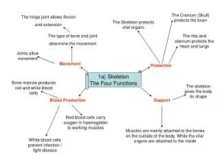
Four General Functions Ingestion Digestion Absorption Excretion
Ingestion Taking food into the body (EATING)
Digestion • The breaking down of food by chemical and mechanical means into usable forms.
Types of Digestion • Mechanical Digestion – Physically breaking down of food into smaller pieces: - Grinding of teeth to soften food - Churning of food by smooth muscles to mix with digestive enzymes • Chemical Digestion - series of catabolic reactions that break down large molecules into smaller molecules.
Absorption The passage of digested food from the alimentary canal into the cardiovascular and lymphatic systems for transportation to body cells
Defecation (Excretion) The elimination of indigestible substances from the alimentary canal
Gastrointestinal Tract (Alimentary Canal) • A continuous tube extending from the mouth to the anus • Organs of the Alimentary Canal • mouth - pharynx - esophagus • stomach • small intestine - large intestine
Accessory Organs Organs that help with digestion by secreting chemicals such as enzymes and bile. • salivary glands (3 pairs. Parotid glands near the ears. • liver • gallbladder - pancreas
Saliva and Salivary Amylase Salivary glands produce saliva which includes water, mucin, bicarbonate ions, and the enzyme, salivary amylase. Salivary amylase helps to chemically break down starch into the disaccharide maltose.
Mouth(Oral or Buccal Cavity) • Cheeks • Lips • Hard Palate • Soft Palate • Uvula • Tongue • Papillae • Lingual Frenulum
Teeth • Accessory structures of the digestive system • Deciduous teeth (baby teeth) - 20 • Permanent teeth - 32 • Incisors (8) - 4 on top, 4 on bottom • chisel shaped - front of mouth • Canines (4) - 2 on top, 2 on bottom • sharp pointed tearing teeth • Premolars (8) - 4 on top, 4 on bottom • Molars (12) - 6 on top, 6 on bottom • broad, flat, crushing teeth
Anatomy of a Tooth • Crown - exposed portion of the tooth above the gum line • Neck - constricted junction line in the tooth between the crown and the root • Root - one to three projections of the tooth that are embedded in the sockets of the alveolar processes of the mandible and maxillae
Anatomy of a Tooth Gingiva – the gums or epithelial tissue that help to protect the root of the tooth. Periodontal Ligament - dense fibrous connective tissue attached to the socket walls and the cemental surface of the roots of the teeth to anchor the teeth. Serves as a shock absorber when chewing
Anatomy of a Tooth • Enamel - outermost portion of the tooth, protects the tooth from wear and tear • the hardest substance in the body • Dentin - calcified connective tissue that gives the tooth its basic shape and rigidity • Pulp – fleshy part of the tooth that contains the blood vessels and nerves. • Cementum - a bone-like substance that covers the dentin of the root
Anatomy of a Tooth • Root Canal – the openings in the root that allows for the passage of blood vessels and nerves.
Digestion in the Mouth • Mechanical Digestion • Chewing (Mastication) • Tongue manipulates the food • Teeth grind up the food and mix it with saliva • The result of mechanical digestion is a soft flexible mass of food called a bolus • Chemical Digestion • Salivary amylase initiates the breakdown of carbohydrates • Only chemical digestion in the mouth
Processes of the Digstive System Deglutition – swallowing
Processes of the Digestive System • Mastication – chewing • Movement of skeletal muscles to close the mandible or jaw to help move the teeth break down food.
Maceration • The breaking down of food into a liquid paste called chyme. Occurs in the stomach.
Segmentation • Short muscle contractions to mix the chyme with enzymes and other secretions. Some propulsion of the chyme also occurs.
Peristalsis • Involuntary smooth muscle contractions to propel food/chyme/wastes through the alimentary canal.
Haustral Churning • Slow, segmental churning within the large intestinal pouches (haustra) which helps to facilitate water reabsorption.
Pharynx • Also called the throat. • Serves as a passageway for food and air. • Also helps in the formation of words.
Esophagus • About 10 inches long • Does not participate in digestive processes – transportation of the bolus to the stomach by peristalsis. • Moves food down into the stomach • Esophageal hiatus - opening in the diaphragm for the esophagus • Joins the stomach at the cardiac opening.
Stomach • J-shaped enlargement of the digestive tract located just below the diaphragm • Superior portion - continuation of the esophagus • Inferior portion empties into the duodenum - pylorus • Position and size of the stomach varies from individual to individual
Histology of the Stomach • Composed of the same four tissue types as the other structures of the alimentary canal • When the stomach is empty the mucosa lie in large folds called rugae. • Mucosa contains gastric glands to make gastric juice. • Chief Cells • Parietal Cells • Goblet Cells
Gastric Glands • Parietal Cells • Secrete hydrochloric acid to activate pepsinogen. • Goblet Cells • Produce mucous for protection • Chief Cells • Secrete Pepsinogen • Enzyme to digests proteins to dipeptides
Anatomy of the Stomach • Fundus • The upper, rounded part of the stomach that will expand to hold more food. • Body • The major portion of the stomach. • Pylorus • The distal, narrow end of the stomach that connects to the small intestine
Anatomy of the Stomach, p.2 • Rugae • Fold of the stomach that allow for stretching. • Cardiac Opening • The opening into the stomach. Is not a true sphinteras food can flow into and out of the stomach. • Pyloric Sphincter • The muscle in the pylorus that controls the movement of chyme into the duodenum (first part of the small intestine)
Mechanical Digestion in the Stomach • Several minutes after food enters, the stomach generates mixing waves that churns the food inside - maceration • Food mixes with gastric juices and is converted into a thin liquid called chyme
Absorption in the Stomach • Does not participate in the absorption of food molecules into the blood • However, can absorb some substances through the stomach wall • Water • Weak glucose concentrations • Electrolytes • Certain drugs (aspirin) • Alcohol
Chemical Digestion in the Stomach • Cephalic Phase - reflexes initiated by sensory receptors in the head • sight - smell - taste • thought of food • Gastric Phase - sensory receptors in the alimentary canal and stomach initiate nervous and hormonal chemical digestive processes • Intestinal Phase - secretion of stomach enzymes that removes nutrients from food
Pancreas • Oblong gland that lies posterior to the greater curvature of the stomach • Connected by the pancreatic duct to the duodenum • Composed of clusters of glandular epithelial cells. • Pancreas is the only organ classified as both an endocrine AND an exocrine gland.
Pancreas • Two main types of Pancreatic Cells: • Pancreatic Islets-Islets of Langerhans (1%) • Hormones: insulin, glucagon, somatostatin • Acini Cells (99%) • Digestive pancreatic enzymes
Pancreatic Juice • Alkaline mixture of fluid and digestive enzymes from the acini cells • Pancreatic digestive enzymes: • Pancreatic amylase - carbohydrate digestion • Pancreatic lipase - fat digestion • Chymotrypsin-Trypsin-Carboxypeptidase - protein digestion • Nucleases - nucleic acid digestion • Regulated by the intestinal hormones secretin and cholecystokinin