Download
1 / 69
811 likes | 1.23k Views
UNIT 4: ANIMAL TISSUE. Campbell and Reece, 2010. Chapter 40 p. 856 – 860. WHERE DO TISSUES FIT IN THE LIVING ORGANISM?. Life begins as a single cell called a zygote after fertilization This cell differentiates into a variety of tissues. (Group of cells with the same function)

E N D
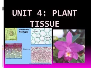
UNIT 4: ANIMAL TISSUE • Campbell and Reece, 2010. Chapter 40 p. 856 – 860
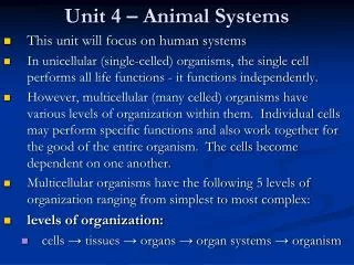
WHERE DO TISSUES FIT IN THE LIVING ORGANISM? • Life begins as a single cell called a zygote after fertilization • This cell differentiates into a variety of tissues. (Group of cells with the same function) • A group of tissues with the same function forms an organ. • Group of organs – Organ system • Group of organ systems - Organism
WHAT IS TISSUE? • SPECIALIZED CELL of the same type that perform a common function in the body.
MAJOR TYPES OF ANIMAL TISSUE • EPITHELIAL TISSUE: Covers or lines body cavity and forms glands. • CONNECTIVE TISSUE: Binds different organs together and support the body. • MUSCLE TISSUE: Movement • NERVOUS TISSUE: Senses and transmits impulses
SQUAMOUS EPITHELIAL TISSUE STRUCTURE • Single layer thin, large, flattened cells • Cells have irregular shape and are closely packed in mosaic form. • Cells are attached to a basement membrane. FUNCTIONS • Diffusion of gasses can occur easily over surface. • Supply friction free surface for blood flow. • Protects and supports underlying tissue
SQUAMOUS EPITELIAL TISSUE WHERE? Lining of lungs and blood vessels In mouth and esophagus.
CUBOIDAL EPITHELIAL TISSUE STRUCTUTE • Square cells, closely packed in a single layer. • Cells attached to a basement membrane. FUNCTION • Absorbs molecules
CUBOIDAL EPITHELIAL CELLS WHERE? Lining of kidney tubules Lining various glands.
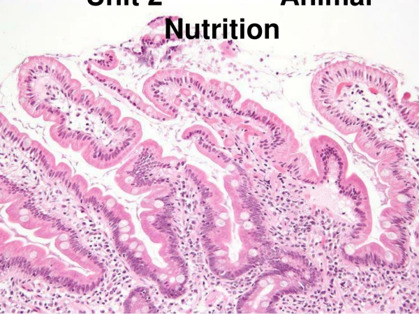
COLUMNAR EPITHELIAL TISSUE STRUCTURE • Tall elongated, column-shaped cells. • Nucleus near the base of the cell. • Cells are attached to a basement membrane. FUNCTIONS • Plays a role in absorption of nutrients. • Protects and supports underlying tissues.
COLUMNAR EPITHELIAL TISSUE WHERE? Lining of small intestine, stomach and oviducts
CILLIATED CULUMNAR EPITHELIAL TISSUE STRUCTURE • Comprises of columnar cells with cilia at their free edges FUNCTIONS • Sweeps impurities towards throat • Improves movement of substance through the ducts.
CILLIATED COLUMNAR EPITHELIAL TISSUE WHERE? Lining of trachea and nostrils, bronchi and sperm ducts)
GLANDULAR COLUMNAR EPITHELIAL TISSUE STRUCTURE • Consist of cuboidal and columnar epithelial cells. • Unicellular glandular cells with goblet cells scattered among non-glandular cells. FUNCTIONS • Produce mucus which serves to lubricate the intestine and make movement of food easier. • Keep respiratory passage moist. • Secrete enzymes, hormones, sweat, wax and saliva.
GLANDULAR COLUMNAR EPITHELIAL TISSUE WHERE? In small intestine and respirative passages. Occur in salivary glands, thyroid gland and sebaceous glands in the skin.
CONNECTIVE TISSUE FIBROUS CONNECTIVE TISSUE SUPPORTIVE CONNECTIVE TISSUE FLUID CONNECTIVE TISSUE
FIBROUS CONNESTIVE TISSUE LOOSE AREOLAR FIBROUS CONNECTIVE TISSUE DENSE WHITE FIBROUS CONNECTIVE TISSUE DENSE YELLOW FIBROUS CONNECTIVE TISSUE ADIPOSE TISSUE
LOOSE AREOLAR FIBROUS CONNECTIVE TISSUE • Occurs beneath the skin and most epithelial layers connecting organs together. • Filling the spaces between organs and muscles, around muscles and blood vessels
DESCRIPTION OF AREOLAR- • Loosely arranged tissue • In jelly-like matrix there are 4 types of living cells and two types of non-living cells. • Macrophages • Mast cells • Fat cells • Fibroblasts • Collagen fibres • Elastic fibres
FUNCTIONS OF AREOLAR- • Areolar tissue binds organs or organ components together and supports structures. • Allows for movement between structures. • The matrix allows for diffusion of substances like gases, nutrients, hormones and wastes to and from the blood.
Fibroblasts – secrete collagen and produce elastic fibres. • Macrophages – Engulf foreign particles by means of phagocytosis to defend the body against infection. • Mast cells – They secrete the matrix, release histamine during inflammation, produce heparin (anticoagulant). • Fat cells – Synthesis and storage of fats. • Collagen fibres strengthen the tissue. • Elastic fibres – Allows for expansion and contraction.
DENDE WHITE FIBROUS CONNECTIVE TISSUE • In dermis of skin, tendons FUNCTIONS: • Attach muscle to bones
DESCRIPTION OF DENSE WHITE- • Consist of mainly white collagen non-elastic fibres. • The collagen fibres are non-elastic, densely packed, arranged in parallel bundles, and has a shiny appearance. • Fibroblasts are elongated cells and occur parallel in rows. • Minimum amount of matrix.
YELLOW FIBROUS CONNECTIVE TISSUE • In ligaments FUNCTIONS: • Attach bone to bone • Hold bones of a joint in place. • Restrict movement of bones and prevent dislocation during normal movement.
DESCRIPTION OF DENSE YELLOW- • Consist of white collagen fibres and a network of yellow elastic fibres. • Ligaments are strong and flexible.
ADDIPOSE TISSUE • Found beneath the skin, around heart and other organs. FUNCTION: • Insulation • Stores fat. DESCRIPTION: • Large round cells filled with fat or oil.
SUPPORTIVE FIBROUS CONNECTIVE TISSUE HYALINE CARTILAGE WHITE FIBROUS CARTILAGE YELLOW ELASTIC CARTILAGE BONE
HYALINE CARTILAGE • Occurs at the ends of bones in movable joints. Parts of the larynx. • In the walls of the trachea and bronchi. • In between the ribs and the sternum. • On the tip of the nose
DESCRIPTION • Contains fine collagen fibres. • White translucent matrix. • Cartilage tissue is surrounded by a fibrous capsule called the perichondrium with blood vessels for nutrition. • It consists of living cells called chondrocytes. • They occur in small fluid-filled spaces called lacunae. In the lacunae the cells are arranged singular or in groups.
FUNCTIONS • Chondrocytes produce a rubber matrix called chondrin. • Reduce friction at joints • Attach bones firmly to other bones. • Keeps tubes open (C-shaped in trachea). • Forms permanent structures (nose). • Longitudinal growth of long bones.
FIBROCARTILAGE • Occurs as disks between the vertebrae. • Surrounds the edges of the sockets of ball-and-socket joints. • Between the pubic bones in front of the pelvic girdle.
FUNCTIONS • It serves as shock absorbers between adjacent vertebrae. • It deepens sockets to make dislocation less easy.
DESCRIPTION • Contain a large number of collagen fibres. • White translucent matrix. • Cartilage tissue is surrounded by a fibrous capsule called the perichondrium with blood vessels for nutrition. • It consists of living cells called chondrocytes. • They occur in small fluid-filled spaces called lacunae. In the lacunae the cells are arranged singular or in groups.
ELASTIC CARTILAGE • In the ear lobe • At the tip of the nose • In the epiglottis. • Septum of nose
FUNCTIONS • It maintains the shape and flexibility of the ear lobe and tip of the nose. • It strengthens and supports the ear, nose and epiglottis.
DESCRIPTION • Contain collagen fibres and a large network of elastic fibres. • Translucent matrix. • Cartilage tissue is surrounded by a fibrous capsule called the perichondrium with blood vessels for nutrition. • It consists of living cells called chondrocytes. • They occur in small fluid-filled spaces called lacunae. In the lacunae the cells are arranged singular or in groups.
COMPACT BONE • Occur in the bones of the skeleton
FUNCTIONS • Support • Protection • Locomotion • Red blood corpuscles and white blood cells are produced in the red bone marrow.
DESCRIPTION • The matrix of compact bone consists of collagen fibrestogether with calcium, phosphorus and magnesium. • These components are arranged in circles called lamellae, around the Haversian canal, containing artery, vein and nerve. • Contain bone cells called osteocytes
FLUID CONNECTIVE TISSUE BLOOD BLOOD PLASMA ERYTHROCYTES LEUCOCYTES BLOOD PLATELETS
BLOODPLASMA WHERE? • Matrix in which the blood cells are found. FUNCTIONS: • Transport various formed elements to body parts. • Dispersion medium. • Transports digested food. • Transports cellular waste. • Transports hormones. • Plays a role in regulation of body temperature. • Transports antibodies DESCRIPTION • Straw color fluid consisting mainly of water with many substances in solution like nutrients, organic waste, inorganic salts and ions, plasma protein, dissolved gases, hormones, enzymes and antibodies.
ERYTHROCYTES (RED BLOOD CELLS) FUNCTIONS: • Transports oxygen in the blood from the lungs to all living cells. • Transports some of the carbon dioxide from the body tissues the lungs. • Play a role in the clotting of blood. • Play a role in the regulating of pH of body fluids
DESCRIPTION • Small round biconcave disks filled with cytoplasm, but without a nucleus. • Contain the red pigment hemoglobin. • Hemoglobin contains 4 units, each with the protein globin and a complex iron-containing, structure called heme. The iron form a loose association with oxygen and in this way red blood cells transport oxygen and readily give it up to tissues.
LEUCOCYTES(WHITE BLOOD CELLS) FUNCTIONS: • Engulf infections by means of phagocytosis. • Produce antibodies. • Defense or immunity of body
DESCRIPTION • Larger cells with nuclei. • Without staining they appear transparent. • Irregular is shape. – there are 5 types of leucocytes based on differences in size and nuclear shape: • Neutrophils • Eosinophils • Basophils • Monocytes. • Lymphocytes.