Download
1 / 62
850 likes | 2.42k Views
Pulse- Oximetry. Presented By Dr Satish Negi Moderated By Dr Manoj Panwar. Defination. A non invasive technology to monitor oxygen saturation of haemoglobin. History. In early 1940.Glen Malkikan coined the term oximeter . . MATHEES- father of oximetry

E N D
Pulse-Oximetry Presented By Dr SatishNegi Moderated By Dr ManojPanwar
Defination • A non invasive technology to monitor oxygen saturation of haemoglobin.
History • In early 1940.Glen Malkikan coined the term oximeter. • MATHEES- father of oximetry • 20 papers in1934 –1944 • HERTZMAN 1937 –use of photoelectric finger plethsmography • 1975 –concept of pulse oximetry –Japan • In 2008 modification continued and term High Resolution Pulse Oximetry come into existence
Introduction • Also called the fifth vital sign • low SpO2 provide warning of hypoxemia before other signs such as cyanosis or a change in heart rate are observed. • Until the 1980s, noninvasive oximeters, known as ear oximeters, were large, expensive, and cumbersome.They required “arterialization” by heat or chemical treatment, and their utility was limited by difficulties in differentiating light absorbance of arterial blood from that of venous blood and tissues. • Technical advances, including light-emitting diodes (LEDs), miniaturized photodetectors, and microprocessors, allowed the creation of a new generation of oximeters, which were smaller, less expensive, and easier to use. • These differentiate the absorption of light by the pulsatile arterial component from the static components, so they are called pulse oximeters
Oxygen Saturation • Saturation is defined as ratio of O2 content to oxygen capacity of Hb - expressed as percentage. • Desaturation leads to Hypoxemia – a relative deficiency of O2 in arterial blood. (PaO2 < 80mmHg – hypoxemia)
Oxygen saturation will not decrease until PaO2 is below 85mmHg. • At SaO2 of 90%, PaO2 is already 60mmHg. • Rough guide for PaO2 between saturation of 90%-75% is PaO2 = SaO2 - 30. • (SaO2< than 76% is life threatening.)
Fractional Saturation • This is Ratio of oxygenated Haemoglobin to sum of all haemoglobin in blood. Fractional saturation = HbO2 - -------------------------- HbO2+ Hb+ Met Hb +CO Hb
Functional Saturation • This is a measure of ratio between HbO2 and sum of oxygenated and reduced Hb. • Functional Saturation= HbO2 ------------ HbO2 + Hb
Cont.. • These must be determined by using an in vitro oximeter. • For patients with low dyshemoglobin levels, difference between fractional and functional saturation is very small. • when dyshemoglobin levels are elevated, two values can vary greatly, and pulse oximeter readings may not agree with either the true fractional or functional saturation values.
PRINCIPLES • All atom and molecules absorbed specific wavelength of light.this property is the basis for an optical technique known as spectrophotometry. • Beer Lambert Law • Lambert’s Law- states that when a light falls on a homogenous substance,intensity of transmitted light decreases as the distance through the substance increase • Beer’s Law- states that when a light is transmitted through a clear substance with a dissolved solute ,the intensity of transmitted light decreases as the concentration of the solute increases
Contd. • Substances have a specific pattern of absorbing specific wavelength – Extinction coefficient Uses two lights of wavelengths • 660nm –deoxyHb absorbs ten times as oxy Hb • 940 nm – absorption of oxyHb is greater • Lab oximeters use 4 wavelengths to measure 4 species of haemoglobin
Optical Recognition of Hemoglobin • Hemoglobin(like all protein)changes its structural configuration when it participate in chemical reaction,and each of the configuration has distinct pattern of light absorption. • Four different forms of hemoglobin represented are :oxygenated hemoglobin(Hbo2),Deoxygenated hemog lobin(Hb),Methemoglobin(MetHb) and Carboxyhemoglobin(CoHb)
Cont.. • Comparing the oxygenated and deoxygenated form of hemoglobin in the Red region of light spectrum(660nm)oxygenated hemoglobin does not absorbed light as wel as deoxygenated Hb.(that’s why oxygenated blood is more intensely red than deoxygenated blood). • While in infrared region(940nm)The opposite is true,and oxygenated Hb absorbed more light effectively than deoxygenatedHb.
Extinction coefficient MetHb Oxy Hb Deoxy Hb COHB 660nm 940nm wavelength
Operating Principles • The pulse oximeter computes the ratio between these two signals and relates this ratio to the arterial oxygen saturation, using an empirical algorithm. • Pulse oximeters discriminate between arterial blood and other components by determining the change in transmitted light caused by the flow of arterial blood
The oximeter pulses the red and infrared LEDs ON and OFF several hundred times per second. • The rapid sampling rate allows recognition of the peak and trough of each pulse wave. • At the trough, the light is transmitted through a vascular bed that contains mainly capillary and venous blood as well as intervening tissue. • At the peak, it shines through all this plus arterial blood. A photodiode collects the transmitted light and converts it into electrical signals. • The emitted signals are then amplified, processed, and displayed on the monitor.
Accuracy • A clinically acceptable level of arterial oxygenation(SaO2 above 70%),the oxygen saturation recorded by pulse oximeters(Spo2) differs by less than 3% from the actual saturation. • pulse oximetry also show a high degree consistency of repeated measurements.
. . Physiology • Oxygen – haemoglobin dissociation curve .
Spo2 100 98 97 96 95 94 93 92 91 90 89 86 83 75 • Pao2 mmhg • 120 • 110 • 92 • 80 • 74 • 69 • 66 • 63 • 60 • 58 • 56 • 51 • 47 • 40
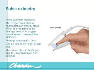
Pulse oximetry uses light to work out oxygen saturation. Light is emitted from light sources which goes across the pulse oximeter probe and reaches the light detector. • If a finger is placed in between the light source and the light detector, the light will now have to pass through the finger to reach the detector. Part of the light will be absorbed by the finger and the part not absorbed reaches the light detector.
The amount of light that is absorbed by the finger depends on many physical properties and these properties are used by the pulse oximeter to calculate the oxygen saturation. • The amount of light absorbed depends on the following: • 1. concentration of the light absorbing substance. • 2. length of the light path in the absorbing substance • 3. oxyhemoglobin and deoxyhemoglobin absorbs red and infrared light differently
Type of Oximetry • Transmission Pulse Oximetry • light beam is transmitted through a vascular bed and is detected on opposite side of that bed. • Reflectance Pulse Oximetry • relies on light that is reflected to determine oxygen saturation. The probe has both an LED and a photodiode. advantage - its signal in low perfusion is better. limitations -The probe design must eliminate light that is passed directly to the probe or is scattered in the outer surface of the skin. The signals are weaker than those found in transmission. this interfere in noisy enviorement and it is costly.
PLETHYSMOGRAPHY • Pulse oximeters show pulsatile change in absorbance in a graphical form. This is called the "plethysmographic trace " or "pleth“ • It tells you how good the pulsatile signal is. • If quality of pulsatile signal is poor- then calculation of oxygen saturation may be wrong. • The pulse oximeter uses very complicated calculations to work out oxygen saturation
Equipment • Probes • The probe (sensor, transducer) is the part that comes in contact with the patient. • It contains one or more LEDs (photodiodes) that emit light at specific wavelengths and a photodetector (photocell, transducer). • The LEDs provide monochromatic light. This means that they emit a constant wavelength throughout their life, so they never need recalibration. • Probes may be reusable or disposable. They have the same accuracy.
Self-adhesive probes are less susceptible to motion artifact and are less likely to come off if the patient moves than those that clip on. • However, they are usually not as well shielded from ambient light as clip-on probes. • probes are available in different sizes. If a probe is too large for the patient, some of the light output from the LED can reach the photocell without passing through tissue, and falsely high SpO2 readings will be produced . • The photocell may not align with the probe, and readings will not be possible.
To reduce contamination, a glove, the finger of a glove, or other covering may be used either over the application site or over the probe . • Cable • The probe is connected to the oximeter by an electrical cable. Cables from different manufacturers are not interchangeable. • Console • Many different consoles are available . Most oximeters that are used in the operating room are part of a physiologic monitor. Most stand-alone units are line operated but will work on batteries, making them useful during transport.
Most instruments provide an audible tone whose pitch changes with the saturation. operator can be made aware of changes in SpO2 without looking at the oximeter. • Alarms are commonly provided for low and high pulse rates and low and high saturation. Many units generate an alarm when the probe is not properly applied to the patient or if the signal is inadequate. • ASA standards for Basic Anesthetic Monitoring require that the variable pitch pulse tone and low threshold alarm be audible
OximeterStandards • There must be a means to limit the duration of continuous operation at temperatures above 41°C. • The accuracy must be stated over the range of 70% to 100% SpO2. If the manufacturer claims accuracy below 65%, the accuracy must be stated over the additional range. • If the manufacturer claims accuracy during motion, this and the test methods used to establish it must be disclosed in the instructions for use. • If the manufacturer claims accuracy during conditions of low perfusion, this and the test methods used to establish it must be disclosed in the instructions for use.
There must be an indication when the SpO2 or pulse rate data is not current. • If the pulse oximeter is provided with any physiologic alarm, it must be provided with an alarm system that monitors for equipment faults, and there must be an alarm for low SpO2 that is not less than 85% SpO2 in the manufacturer-configured alarm preset. An alarm for high SpO2 is optional. • An indication of signal inadequacy must be provided if the SpO2 or pulse rate value displayed is potentially incorrect.
Any discoloration of the nail bed can affect the transmission of light through the digit. • dark nail polish, such as blue, green, brown, or black colors, and bruising under the nail can limit the transmission of light and result in an artificially decreased Spo2 value. • If the nail polish cannot be removed, the sensor can be placed in a lateral side-to-side position on the finger to obtain readings if no other method of sampling the arterial bed is available
Sites of probe placement • A disadvantage of placing a probe on an extremity is that detection of desaturation and resaturation is slower than when probes are placed more centrally . Response time may be quicker when the probe is placed on the thumb. • Motion artifacts are less frequent when the probe is placed on one of the larger fingers. • The probe should not be on the index finger during recovery. As a patient awakens, the patient often will want to rub his or her eye, usually with the index finger. If the oximeter probe is on that finger, the cornea can be scratched. • In general, the arm opposite from that on which the blood pressure cuff is applied or in which an arterial catheter has been inserted should be used.
The pulse oximeter is sometimes integrated with the noninvasive blood pressure monitor so that the pulse oximeter will not alarm during the inflation cycle if placed on the same arm as the blood pressure cuff. • Occasionally, poor function may occur with probe attachment to the same extremity as the intravenous infusion, due to local hypothermia and vasoconstriction • Toe Detection of desaturation or resaturation will not be as rapid as with more centrally placed probes .
The toe may provide a more reliable signal in patients who have had an epidural block . • Nose • Nasal probes respond more rapidly to changes in saturation than probes placed on extremities. The bridge, the wings of the nostrils, and the nasal septum can be used • It has been recommended under conditions such as hypothermia, hypotension, and infusion of vasoconstrictor drugs. • If the patient is placed in the Trendelenberg position, venous congestion may occur around the nose, causing the pulse oximeter to display artificially low saturations
Ear • The earlobe should be massaged for 30 to 45 seconds with alcohol or vasodilator or EMLA cream can be applied for 30 minutes prior to probe application to increase perfusion. • useful when there is finger motion. • A steep head down position may result in erroneous readings. • Tongue • A tongue probe can be made by placing a malleable aluminum strip behind the probe to allow it to bend around the tongue. • This site may be especially useful in patients who have burns over a large percentage of their body surface . • Desaturation and resaturation is detected by a probe at the tongue quicker than one on the finger or toe.
A lingual probe is more resistant to signal interference from electrosurgery than probes placed on peripheral sites. • Cheek • A probe with a metal strip backing can be used to hold a disposable probe around the cheek or lips. • Buccal pulse oximetry is more accurate than finger pulse oximetry. • effective during hypothermia, decreased cardiac output, increased systemic vascular resistance, and other low pulse pressure states. • Esophagus • This probe uses reflectance oximetry. The esophagus, a core organ, is better perfused than the extremities during states of poor peripheral perfusion
It reflects changes in arterial saturation more quickly than peripheral sites such as the finger. • useful for patients who have extensive burns • Forehead • A flat reflectance pulse oximeter sensor can be used on the forehead. Pressure on the probe from a headband or pressure dressing may improve the signal • The forehead is less affected by vasoconstriction from cold or poor perfusion than the ear or finger. • Changes in saturation can be detected more rapidly at the forehead than at the finger. However, pooling of venous blood due to compromised return to the heart may cause low saturation readings in supine patients
Pharyngeal pulse oximetry by using a pulse oximeter attached to a laryngeal mask may be useful in patients with poor peripheral perfusion. • Flexible probes may work through the palm, foot, penis, ankle, lower calf, or even the arm in infants • Pulse oximetry may be used to monitor fetal oxygenation during labor by attaching a reflectance pulse oximetry probe to the presenting part . • A disadvantage is that the probe has to be placed blindly and may be positioned over a subcutaneous vein or artery, which will affect the reliability of the readings.
uses • Monitoring oxygenation: PACU,transport. • Detect inadvertent bronchial intubation • Managing one lung anaesthesia • Weaning from artificial ventilation • Controlling 02 administration(lowest safe flow and concentration) • Monitoring peripheral circulation(distal to fracture) but not helpful in warning compartment syndrome. • Evaluation of sympathetic block
SBP measurement • Locating vessels(axillary artery) • Avoiding hyperoxemia • Monitoring hypovolemia(skipping beats) • Monitoring sympathetic tone(dicrotic notch) • Pulse rate • Arrhythmias • pulse oximetry can balance blood gas analysis • neonatal care(one element of sugesting congenital heart ds.) • During thoracic anaesthesia
Limitations and Disadvantages • Failure to Determine the Oxygen Saturation • Factors that are reported to contribute to higher failure rates include ASA physical status 3, 4, or 5 patients, orthopedic, vascular, and cardiac surgery; electrosurgery use; hypothermia; hypotension; low hematocrit; and motion • Poor Function with Poor Perfusion • Pulse oximeters require adequate pulsations to distinguish light absorbed from arterial blood from venous blood. • Readings may be unreliable or unavailable if there is loss or diminution of the peripheral pulse (proximal blood pressure cuff inflation, external pressure, improper positioning, hypotension,hypothermia, Raynaud's phenomenon, low cardiac output, hypovolemia, peripheral vascular disease
Erratic Performance with Dysrhythmias • double- or triple-peaked arterial pressure waveform that confuses the pulse oximeter, so it may not provide a reading • Different Hemoglobins • Most pulse oximeters are designed to detect only two species of hemogobin: reduced and oxygenated. Whole blood often contains other moieties such as carboxyhemoglobin, sulfhemoglobin, and methemoglobin. • Methemoglobin • Normally less than 1% of the total hemoglobin, methemoglobin (metHb) is an oxidation product of hemoglobin that forms a reversible complex with oxygen and impairs the unloading of oxygen to tissues. • Methemoglobin absorbs light equally at the red and infrared wavelengths that are used by most pulse oximeters. When compared with functional saturation, most pulse oximeters give falsely low readings for saturations above 85% and falsely high values for saturations below 85% .
Carboxyhemoglobin • Carboxyhemoglobin has an absorption spectrum similar to that of oxyhemoglobin at 660nm, so most pulse oximeters will over-read SpO2 by the percentage of carboxyhemoglobin present • An increase in HbCO may occur during laser surgery in the airway, but the levels are not high enough to keep pulse oximetry from reliably estimating saturation . • Pulse oximeters that differentiate between oxyhemoglobin and carboxyhemoglobin are now available • Fetal Hemoglobin fetal hemoglobin (Hb F) does not affect the accuracy of pulse oximetry to a clinically important degree
Sulfhemoglobin • Sulfhemoglobinemia may be caused by drugs such as metoclopramide, phenacetin, dapsone,and sulfonamides. Sulfhemoglobin causes the pulse oximeter to display artifactually low oxygen saturation. • Bilirubinemia • Severe hyperbilirubinemia can cause an artifactual elevation of metHb and carboxyhemoglobin when using in vitro oximetry but does not affect pulse oximetry readings. • Low Saturations • Pulse oximetry becomes less accurate at low oxygen saturations . This inaccuracy is greater in patients with dark skin. It should be used with caution in patients with cyanotic heart disease. • Malpositioned Probe