Download
1 / 53
551 likes | 826 Views
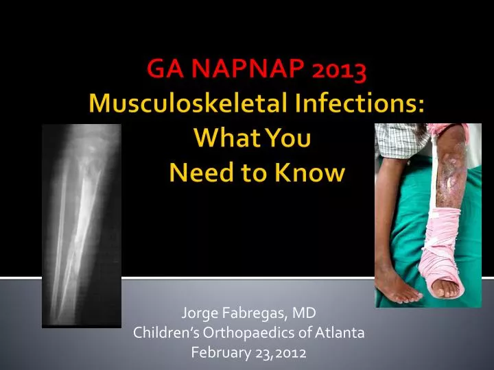
Jorge Fabregas, MD Children’s Orthopaedics of Atlanta February 23,2012. GA NAPNAP 2013 Musculoskeletal Infections: What You Need to Know. Goals. Understand evaluation of patient with possible infection. Incidence Prevalence Etiology Treatment Septic Arthritis Osteomyelitis

E N D
Jorge Fabregas, MD Children’s Orthopaedics of Atlanta February 23,2012 GA NAPNAP 2013Musculoskeletal Infections: What You Need to Know
Goals Understand evaluation of patient with possible infection Incidence Prevalence Etiology Treatment • Septic Arthritis • Osteomyelitis • Soft Tissue Infections
The Great Imitator • What defines infection? • Fever • Pain • Swelling • Warmth • Irritable joint • Pus • Wound drainage • ESR, CRP, WBC • Aspiration • cell count, diff, gram stain • Radiographic changes • Positive culture • 20% no organism identified Floyed and Steele 2003 • Positive blood culture • Response to antibiotics • Absence of other pathology
2yF refusing to walk • Pain x 24 hours • Left sided limping, then inability to bear weight • Crying, ill-appearing • Family brings to ED for evaluation
History • No trauma • Possible fever • Low appetite • Upper respiratory infection 2 weeks ago • no antibiotics • No sick contacts • Goes to daycare • No PMH/PSH
Physical examination • 37.2, 131, 30, 97/72, 95% RA, 11.1kg • Ill-appearing • Laying still • Left hip flexed, abducted, externally rotated • Left hip irritable • No pain ROM knee or ankle • No tenderness knee and distal • Wiggles toes • Neurovascularly intact
Laboratory evaluation • CBC • WBC 10.36, 63% PMNs • Hgb 12.4 • Plt 296 • ESR • 15 • CRP • 7.9 • Blood cultures
Imaging • Xray • normal • Ultrasound • effusion • MRI • Effusion • No osteo • No abscess • Perfusion
Treatment • OR for aspiration and I&D left hip • Small amount of viscous, cloudy, bloody fluid • Sent for culture and DNA studies • Closed over drain • Antibiotics • ID consult • PICC • Blood and synovial fluid cultures no growth to date
Joint aspiration • Inoculate directly into blood culture bottle to enhance culture of fastidious organisms • K. kingae • WBC > 50,000/mm3 with predominance of neutrophils (75%) consistent with infection • WBC <25, 000 in 34%of patients • WBC can be elevated in JRA • Gram stain positive in 30-50% of patients • Cultures positive in 50-80% of patients • Low protein, high lactate and low glucose levels compared to serum indicative of infection
Kocher criteria JBJS 2003, JBJS 2004 • Fever 38.5 • Refusal to bear weight • ESR 40 mm/hr • Serum WBC >12,000 cells/mm3 • 4 predictors 99.6% (93%) • 3 predictors 93.1% (72.8%) • 2 predictors 40% (35%) • 1 predictor 3% (9.5%) • CRP > 2.0 Caird et al JBJS 2006 • 5 predictors 98% • 4 predictors 93% • 3 predictors 83%
Synovial fluid analysis • Wide range WBC possible, often lower with atypical organisms • Organism identified 30% Lyon and Evanich JPO 1999 • No significant clinical or laboratory differences
Management • Surgical decompression of joint space • Create capsular window to ensure continued drainage • Leave drain in place until drainage decreases significantly • If no rapid improvement of symptoms • Reexploration • Further diagnostic workup
Epidemiology • Incidence 1:5000 Sonnen and Henry 1996 • Acute hematogenous osteomyelitis, age < 13 • Septic arthritis twice as common Gutierrez 1997 • Most common in 1st decade • ½ younger than 5 Gillespie 1987 • Lower extremity 70-90% • Hip 54% Wang 2003 • Incidence decreasing • Awareness, immunization, antibiotics
Acute HematogenousOsteomyelitis & Septic Arthritis • Metaphysis may be within the joint capsule • proximal part of the femur, humerus, ankle and proximal radius. • result in the coexistence of septic arthritis and osteomyelitis
Newborns: infection can cross the physis and enter epiphysis and joint Capillaries on metaphyseal side of growth plate do not cross growth plate after 6 -18 months Acute Hematogenous Osteomyelitis & Septic Arthritis
Clinical Manifestations • Trauma or URI may precede symptoms • Joint pain, fever, irritability, anorexia, limp • Redness, swelling, and warmth over affected joint • Painful restricted ROM • Hip in flexion, abd, ER
Diagnosis • Blood culture positive 30-50% • Peripheral blood • WBC, ESR and CRP elevated • CRP occasionally not elevated, especially with K. kingae • Radiology • Evaluate for other causes: trauma, malignancy, osteomyelitis
Diagnosis • Important to differentiate between septic joint and transient synovitis • Considerable overlap in clinical and lab findings • Hip pain • Refuse to WB, limp • Pseudoparalysis • Hip held in flex, abd, • Recent viral illness • Treatment varies dramatically • NSAID’s vs Open arthrotomy • Predominates in 5-10 year old males • Radiology usually normal • US screening • modality of choice for joint effusion
Most common organisms • Staphylococcus aureus • 70-90% cases musculoskeletal infection Blyth JBJS 2001 • Newborns • S. aureus, Group B strep, Gram negative rods • Children • S. aureus, Group A β-hemolytic strep, Strep pneumo, Kingella kingae, (H. influenza) • Adolescents • Gonococcus • Sickle cell • Salmonella • Foot puncture wound • Pseudomonas
Management • Most antibiotics achieve high synovial fluid concentrations • IV therapy until clinical improvement and CRP returning to normal • Uncomplicated septic joint (no concurrent osteo) • 3-4 days of IV therapy followed by appropriate oral therapy • Duration depends on response to therapy and on suspected organism • S. pneumoniae, K. Kingae, Hib, N. gonorrhhoeae treated for 2-3 weeks • S. aureus or gram-negative enteric bacteria treated 3-4 weeks
CA-MRSA • Young, previously healthy children • Aggressive skin, soft tissue, and bone infection • Risk factors • Antibiotic use within the preceding year, crowded living conditions, compromised skin integrity, participation in team sports. • mecA gene • Resistance to methicillin and other β-lactam antibiotics • Panton-Valentine leukocidin (PVL) • Cytotoxin • Lyses WBCs, promotes tissue necrosis, allows pathway for CA-MRSA to proliferate in the host • Associated with deep-seated and life threatening infections
CA-MRSA Vanderhave et al JPO 2009 • Review of all patients with CA-MRSA infections requiring orthopaedic care • 27 previously healthy children (18 M, 9F) average age 9.3 years (3mo to 17.7 y) • History of minor trauma (n=4) or sports-related injury (n=5) within 1 week of presentation in 9 of 27 patients (33%). • Clinical presentation involved an extremity in 23/27 • 5 upper extremities and 18 lower extremities • 17 had temp > 38.5 at presentation, 6 over 40 • Osteomyelitis 13, pyomyositis 11, septic arthritis 10, soft tissue or subperiosteal abscess 6, multifocal involvement 13
CA-MRSA Vanderhave et al JPO 2009 • 2 patients treated w/ clindamycin developed resistance • Significant long-term sequelae 9 patients (33%) • 4 chronic osteomyelitis requiring surgery 3-12 mo later • 1 fixed elbow contracture in dominant arm • 1 heterotopic ossification around the hip • 1 destruction of hip due to osteo required THA • 1 distal tibial physeal arrest elected amputation for pain and deformity
Articular cartilage destruction • Proteases, peptidases, collagenases released • Leukocytes, synovial cells, cartilage • Break down cellular and extracellular structure of collagen • Loss of glycosaminoglycans – 8 hours • Softens cartilage • Susceptible to increased wear • Once catalytic enzymes released, living bacteria are not necessary for cartilage destruction to continue
Risk Factors for Poor Outcome • Prematurity • Age less than 6 months • Delay in treatment > 4 days • Concurrant osteomyelitis of femur • Septic dislocation of hip joint
Sequelae of SepticArthritis of the Hip 40% hip infections poor results • Partial or complete destruction of the proximal femoral physis • Osteonecrosis of the femoral head • Trochanteric overgrowth • Pseudarthrosis of the femoral neck • Complete dissolution of the femoral neck and head • Progressive limb-length discrepancy • Varus or valgus alignment of the femoral head • Unstable hip articulation • Hip dislocation • Ankylosis of the hip joint
12yF 3 days right ankle pain • Fevers to 102 • Twisted his R ankle last week • Unable to bear weight x 2 days • Seen at urgent care, dx arthralgia, Tylenol #3 • Warts removed from left knee 1 month ago • Cellulitis treated with antibiotics • PMH: twin born 38 weeks via C-section
Exam • 37.7 °C, 101, 18, 104/77, 100% RA, weight 46.9 kg • Ill-appearing • Generalized maculopapular rash • Right foot and ankle swelling, warmth, maculopapular rash • No open wounds • No fluctuance • Tender over ankle, distal tibia, distal fibula • Ankle joint irritable • Sensation intact • DP and PT pulses palpable
Labs • WBC 18.6, 58% PMNs • Hgb 15.8 • Plt 215 • ESR 10 • CRP 23 • Blood culture
Diagnosis • Attempts to obtain culture should be made • Blood and tissue cultures • Blood cultures positive 30-50% • Tissue critical for diagnosis of organism • Culture and histopathology • Inoculation of material directly into aerobic blood culture bottle facilitates isolation of fastidious organisms • Begin empiric therapy for “most likely” organism
Aspiration • Right ankle, tibia, fibula • Point of maximum tenderness • Gross purulence • Gram positive cocci in clusters
Acute Hematogenous Osteomyelitis • Aspiration • Locate point of maximum tenderness & swelling • Usually metaphyseal • 16 or 18 gauge spinal needle to aspirate • Extraperiosteally, subperiosteally, intraosseously. • Positive in 60% cases (Biopsy 90%)
Management • Institution of appropriate antibiotic therapy • Healthy neonate: Group B Streptococcus most common (S. agalactiae) • Oxacillin or cefotaxime • High risk neonate: S. aureus most common • Oxacillin or cefotaxime plus gentamycin • Infants to 3 years: S. aureus, K. kingae • Cefataxime or cetriaxone and PCN for K. kingae • > 3 years: S. aureus • Oxacillin
Acute Hematogenous Osteomyelitis Diagnosis: • clinical findings, and a high index of suspicion essential. • Unexplained bone pain with fever means osteomyelitis until proven otherwise. • onset is usually sudden • 30% to 50% of patients have had a recent or concurrent nonmuscular infection.
Microbiology • S. aureus most common in all age groups • CA-MRSA becoming more common • Infants <2 months • S. agalactiae, Neisseriagonorrhoeae, gram-negative enteric bacteria, Candida • 2 months – 5 years: • S. aureus, S. pyogenes, S. pneumoniae and K. kingae • > 5 years: • S. aureus, S. pyogenes, N. gonorrhoeae
Osteomyelitis - pathophysiology • Metaphysis • Small terminal vessels beneath physis – slow flow • Few phagocytic cells • Endothelial gaps • Rapidly growing long bones • Trauma 30-50% acute hematogenous osteomyelitis • iv S. aureus lead to infection in metaphysis of injured rabbit Morrissy and Haynes JPO 1989
Inflammation • Intramedullary pressure • Communication with subperiostial space • Ischemia/necrosis • “Bone cellulitis” “Bone abscess”Subperiosteal abscess • Sinus tract to skin may form = cloaca (Latin: “sewer”) • Inaccessible to antibiotics • Chronic osteomyelitis
Treatment • ICU admission • Coagulopathy, petechial rash • I&D right fibula, wound vac placement • Repeat I&D, vac placement • Repeat I&D, closure over a drain • Vancomycin, ceftriaxone → clindamycin → oxacillin • ID consult • Blood cultures: MSSA • Fibula aspiration: MSSA • Afebrile, CRP 7.6
Osteomyelitis – Complications/Sequelae • Bone loss • Need for grafting • Fracture • Growth disturbance • Limb length inequality, angular deformity • Chronic osteomyelitis • DVT
Other Imaging Studies • Ultrasound • can detect fluid collections or abscess • periostitis/surface abnormalities
CT scan • Fast but less useful in early stages • Identifies cortical destruction, bony sequestrum, extraosseous abscess or gas • CT Scan is helpful in chronic cases • small areas of osteolysis (sequestra) • foci of gas, minute foreign bodies
Bone scan • Detect specific lesions or multiple lesions • Useful in initial 48-72 hours of symptom onset • May have cold scan initially • Vascular supply to bone is compromised • Decreased uptake of isotope • Tagged WBC scan can increase specificity for infection (80%) • Positive in other illnesses causing increased osteoblastic activity • Malignancy, trauma, cellulitis, postsurgery, arthritis • Preferred test by some pediatric infectious disease experts • Less expensive than MRI • Sedation not necessary • Useful for multifocal or location of infection not obvious
MRI • Most sensitive modality, but not specific • Soft tissue abscess, bone marrow edema, bone destruction • Preferred test for surgical planning • Limitations • Expense • Sedation in young children • Inability to assess whether other bones are affected • Fracture or bone infarction may not be easily distinguished from infection
Optimal imaging strategy for community-acquired Staphylococcus aureus musculoskeletal infections in children. PediatrRadiol. 2008 Aug;38(8):841-7. • Retrospective review of CA-SA osteomyelitis cases since 2001 at Texas Children's Hospital • 199 children with CA-SA osteomyelitis • MRI bone scintigraphy • n=160 n=35 • sensitivity = 98%53% • CONCLUSION: MRI is the preferred imaging modality for the investigation of pediatric CA-SA musculoskeletal infection because it offers superior sensitivity for osteomyelitis compared to bone scintigraphy and detects extraosseous complications that occur in a substantial proportion of patients.
MRI may eliminate unnecessary surgery for children with suspected musculoskeletal infections. Kan, J.H. American Journal of Roentgenology. November, 2008 • Vanderbilt Children’s Hospital in Nashville, Tenn • 130 children with suspected musculoskeletal infections • 34 patients underwent an MRI after diagnostic or therapeutic intervention • 96 patients had an MRI prior to any procedure • 60% of patients had neither septic arthritis nor osteomyelitis • “The majority of the children in the study group had a diagnostic or surgical procedure which could have been avoided with early MRI evaluation.”
Radiology Summary • No radiographic technique can make or exclude diagnosis with certainty • raise/lower suspicion when applied to a specific clinical situation
Soft Tissue Infections • Cellulitis • Diffuse leukocyte inflammation, hyperemia, edema without abscess. • Group A Beta hemolytic Strep or S. aureus • IV or oral abx • Surgical drainage if abscess forms • Puncture wound • S. aureus, Pseudomonas if athletic shoe • Tetanus toxoid • ER or surgical debridement