Download
1 / 1
10 likes | 139 Views
O. O. H. H. H. O. H. O. R. P. O. O. O. R. C. C. O. O. O. O. O. O. H. H. O. H. H. H. H. O. O. H. H. O. O. R. R. P. P. O. O. O. O. O. O. R. R. C. C. C. C. O. O. O. O. O. O. 40. 40. 14. 14. 14. Chol. Chol. Chol. [. [. [. C] . C] .

E N D
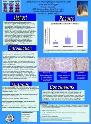
O O H H H O H O R P O O O R C C O O O O O O H H O H H H H O O H H O O R R P P O O O O O O R R C C C C O O O O O O 40 40 14 14 14 Chol Chol Chol [ [ [ C] C] C] 30 30 Cer Cer 14 14 [ [ C] C] 14 14 [ [ C] PE C] PE by bNPC2 by bNPC2 % of transferred lipid % of transferred lipid 20 20 14 14 [ [ C] PC C] PC 14 14 [ [ C] GM3 C] GM3 10 10 14 14 [ [ C] C] Sulfatide Sulfatide 14 14 [ [ C] C] GalCer GalCer 0 0 Adebayo Adebayo et al. et al. 4.2 4.2 5.0 5.0 6.0 6.0 7.4 7.4 pH pH ( ( Unpublished Unpublished ) ) Probing cholesterol homeostasis in model membranes by biotin-streptavidin equilibrium system Ladoke Akintola University of Technology, Ogbomoso, Nigeria Misbaudeen Abdul-Hammed*, a,b,c, Jonathan O. Babalola b, Matthew A. Adebayo b, Bernadette Breiden c, Guenter Schwarzmann c and Konrad Sandhoff c a Department of Pure and Applied Chemistry, Ladoke Akintola University of Technology, Ogbomoso, Nigeria b Department of Chemistry, University of Ibadan, Nigeria c Life and Medical Sciences (LIMES) Institute, c/o Kekulé Institute of Organic Chemistry and Biochemistry, University of Bonn, Federal Republic of Germany University of Ibadan, Nigeria INTRODUCTION Unesterified Cholesterol, together with glycolipids, accumulates in cells leading to Niemann-Pick type C disease, an autosomal recessive neurodegenerative disease 1. Two types of genes, NPC1 and NPC2 responsible for this neurodegenerative disorder have been identified but their respective precise functions are yet to be fully elucidated. NPC1-NPC2 proteins interplay has been elucidated in such a way that NPC2 binds and transfers cholesterol 2-4 to a cholesterol binding site of NPC1 for export out of the endosomal system 5,6. Until now, the study of Cholesterol has been hampered because the lipid lacks a functional group that can be readily tagged without altering its physical and chemical properties. Transfer studies of fluorescent cholesterol esters or cholesterol analogs are biased because of changes in the physicochemical nature of the lipids and might not correctly reflect the properties of unmodified cholesterol. A simple liposomal assay system which allows probing of the intermembrane transfer of radioactive cholesterol from donor to acceptor vesicles is therefore developed as well as a model to measure the vesicle fusion induced by some proteins. Abilities of the lysosomal lipid binding proteins (such as NPC2 protein) to transfer cholesterol in model membranes which mimic the endosomal/lysosomal compartments and to mediate vesicle fusion as well as the influence of lipid compositions were investigated. Other lysosomal lipid binding proteins investigated are saposins A-D and GM2AP while non-lysosomal proteins such as cytochrome C and bovine serum albumin were used as controls. METHODS Fig 2: Model for the transfer and fusion assay. Transfer assay is based on the use of donor liposomes containing [14C]-labelled cholesterol (red) and a biotin-labelled PE (blue), which allows the separation of these membranes by streptavidin-coated magnetic beads. The acceptor liposomes are marked by NBD-PE (green) to control the recovery of these vesicles. After removing the biotin-labelled liposomes, presence of significant radio-labelled cholesterol in the supernatant indicates lipid transfer. Fusion assay is based on the use of donor liposomes containing biotin-labelled PE (blue), and the acceptor liposomes containing both [14C]-cholesterol (red) and fluorescent NBD-PE. Simultaneous decrease in radioactivity and fluorescence in the acceptor vesicle (supernatant) indicates vesicle fusion. Host lipids (PC) are shown in yellow. Acceptor Supernatant Donor LLBP Transfer Assay BioMag Streptavidin Fusion Assay Fused Liposome Fig 1: Model of the roles of NPC1 and NPC2 in cholesterol homeostasis LLBP BioMag Streptavidin Acceptor Donor Intermembrane Transfer of cholesterol mediated by various proteins Fig. 3: (A) Intermembrane transfer of cholesterol mediated by proteins g = glycosylated, u = unglycosylated, Saps A-D = saposins A-D, GM2AP = GM2-activator protein, Mixed protein = a mixture of equimolar Amounts of NPC2,Saps A-D and GM2AP.Only NPC2 significantly transfer cholesterol with bovine NPC2 more effective than its human counterpart.; (B) GM2AP tends to be highly fusogenic protein in the presence of BMP, and separating donor from acceptor vesicles in the presence of GM2AP requires higher amount of streptavidin-coated magnetic beads, which eliminates the pseudo-cholesterol transfer recoreded for the protein in Fig. 3A. This also applies to saposins A, C and D but not NPC2. BMP enhances intermembrane transfer of cholesterol by bovine NPC2 Bis(monoacylglycero)phosphate (BMP), erroneously also called lysobisphosphatidic acid, is a negatively charged lipid which is highly enriched in the internal membranes of the lysosome and required for degradation of small GSL 7. It is formed during the degradation of phosphatidylglycerol and cardiolipin, presumably on the surface of intra-lysosomal vesicles 8,9. In addition to the presence of hydrolyzing enzymes, degradation of intra-lysosomal membranes requires a low pH value, high BMP content, and low cholesterol levels10-11. Fig 4: (A) BMP enhances cholesterol transfer activity of bovine NPC2 protein, with unsaturated one having hígher influence than unnatural saturated BMP; (B) Cholesterol transfer increases with BMP contents at acidic pH B A A B Lipid transfer activity of bovine NPC2 protein is specific to cholesterol and its analogs Cholesterol associates tightly with sphingomyelin in biological membranes via the formation of intermolecular hydrogen bonds and so is ceramide, but the latter forms stronger hydrogen bondswith sphingomyelin thereby making it possible for ceramide to displace cholesterol from mebranes12. Therefore, it is our desire to get more insight into the functional interrelationship of ceramide and cholesterol levels within the internal membranes of endosomes and lysosomes. For this purpose, the ceramide transfer activities of bovine NPC-2 was determined in vitro compared to its transfer activities of cholesterol and other lipids using a recently established assay for the protein-mediated cholesterol transfer between donor- and acceptor liposomes 3. Fig 5: (A) Of all the radiolabelled-lipid probed here, only cholesterol was significantly transferred by bovine NPC2 protein at all the pH investigated. The reaction was carried out at 25°C for 10 minutes using 40 µl of 5 mg/ml streptavidin-coated paramagnetic beads suspension. (B) Ceramide is transferred spnotaneously more than cholesterol and any other lipid under investigation. Vesicle fusion triggered by lysosomal lipid binding proteins Membrane fusion proceeds by an ordered sequence of steps that include the merging of proximal monolayers, stalk formation, generation of hemifusion intermediates and fusion pores opening. Energy (UV lasers), high Ca (2+) concentration, proteins and lipid compositions have been shown to play central roles in cell membrane fusion. Such proteins may include viral proteins (like influenza virus hemagglutinin), SNARES (soluble N-ethylmaleimide-sensitive factor acceptor protein receptor) proteins and some lysosomal proteins, such as saposins C and D. Fig. 6: (A) Vesicle fusion is induced by some lysosomal lipid binding protein at acidic pH (4.2), Protein content = 1.5 µg, 25°C, 10 mins. Naturally occuring BMP catalyzes fusion more than unnatural BMP and ceramide; (B) The fusogenic activity of saposin C decreases with pH (C) NPC2 protein also catalyzes fusion at acidic pH. A pH 4.2 A B C B • ONGOING/ FUTURE RESEARCH PLAN • To investigate how various concentartions of BMP, ceramide and sphingomyelin in the liposomes influence the cholesterol transfer activity of bovine NPC2 at endosomal pH; • To probe the influence of variable BMP, ceramide and sphingomyelin contents in the liposomes on the fusogenic activities of the proteins; • To compare cholesterol transfer activities of bNPC2 and β-cyclodextrin; • To analyze the extent to which highly unsaturated naturally-occuring BMP (e.g. BMP(24:6))catalyzes cholesterol transfer and fusogenic activities of the proteins under consideration • SUMMARY • Of all the lysosomal lipid binding proteins probed in the transfer assay, only NPC2 significantly transfer cholesterol at acidic pH, with bovine NPC2 having higher cholesterol transfer activity more than its human counterpart (Fig. 3) • BMP (a unique negatively-charged phospholipid) mediates the transfer of cholesterol by bovine NPC2 and the cholesterol transfer is pH dependent (Fig. 4) • bNPC2 specifically transfer cholesterol (or some other sterol family member) but not any other lipid under consideration (Fig 5). • Sap C, GM2AP, bNPC2 and some other lysosomal lipid binding proteins mediate vesicle fusion and the fusogenic properties of the proteins were catalyzed by BMP and ceramide (though to a little extent) at acidic pH (Fig 6) ACKNOWLEDGEMENT This work was supported by Deutsche Forschungsgemeinschaft (SFB 645) and by the seventh framework program of the EU-funded “LipidomicNet” (proposal number 202272). • REFERENCES • Pentchev, P.G., Comly, M.E., Kruth, H.S., Tokoro, T., Butler, J., Sokol, J., Filling Katz, M., Quirt, K. M., Marshall, D. C., Patel, S., Vanier, M. T. and Brady, R.O. (1987). FASEB J. 1, 40-45. • Cheruku, S.R., Xu, Z., Dutia, R., Lobel, P. and Storch, J. (2006). J. Biol. Chem. 281, 31594-31604. • 3. Babalola, J.O., Wendeler, M., Breiden, B., Arenz, C., Schwarzmann, G.,Locatelli-Hoops, S. and Sandhoff, K. (2007). Biol. Chem. 388, 617-626. • 4. Xu, Z., Farver, W., Kodukula, S. and Storch, J. (2008). Biochemistry 47, 11134-11143. • 5. Subramanian, K. and Balch, W.E. (2008). PNAS 105(40), 15223-15224. • 6. Infante, R.E., Wang, M.L., Radhakrishnan, A., Kwon, H.J., Brown, M.S. and Goldstein, J.L. (2008). PNAS 105(40), 15287-15292. • 7. W.Möbius, E. van Donselaar, Y. Ohno-Iwashita, Y. Shimada, H.F. Heijnen, J.W. Slot, and H.J. Geuze, Traffic 4 (2003) 222–231 • 8. B. Amidon, A. Brown and M. Waite, Biochemistry 35 (1996) 13995–14002. • 9. J. Brotherus and O. Renkonen, J. Lipid Res. 18 (1977) 191–202. • 10. H. Schulze, T. Kolter and K. Sandhoff (2009). Biochimica et Biophysica Acta, 1793(4) : 674-683. • 11. T. Kolter and K. Sandhoff. (2005) Annu. Rev. Cell Dev. Biol. 21; 81–103. • 12. Megha and E. London (2004) J. Biol. Chem. 279:9997-10004.