Download
1 / 19
220 likes | 605 Views
Baermann’s Apuspara. ZOO515 1433-1434. Baermann technique: Purpose. The Baermann technique is used to separate larvae from faecal material. For example: Diagnosing lungworm infection The identification of third stage larvae [L 3 ] from a faecal culture .

E N D
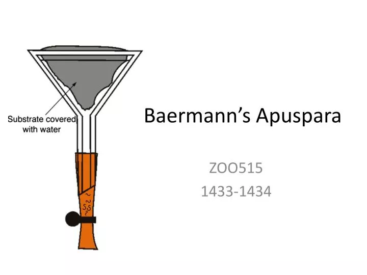
Baermann’sApuspara ZOO515 1433-1434
Baermann technique: Purpose • The Baermann technique is used to separate larvae from faecal material. • For example: • Diagnosing lungworm infection • The identification of third stage larvae [L3] from a faecal culture.
Baermann technique: Principle The Baermann technique is based on the active migration or movement of larvae. Faeces are suspended in water. The larvae move into the water. They sink to the bottom and can be collected for identification.
There is no standard equipment for this technique. Each laboratory adapts its own procedure. Commonly used equipment is illustrated and a list is shown below.
Procedure Take a funnel and fit a short piece of tubing to the stem. Close the tubing with a clamp or spring clip. Support the funnel on a single stand. If more than one sample requires processing at the same time use a plank in which holes have been made to support several funnels.
Procedure Place a double layer of cheesecloth or dental napkin on a disposable paper towel or equivalent on the bench. Using a spoon or spatula weigh or measure approximately 5-10 grams of faecal material. Place the faecal material in the centre of the cheesecloth.
Procedure Form a pouch containing the faecal material by holding the four corners of the cheesecloth together and moulding the cloth around the faecal material.
Procedure Using a rubber band or length of string close the cheesecloth pouch. Push the stick or short metal rod under the rubber band or string so that the pouch can be suspended.
Procedure Place the pouch containing the faecal material in the funnel. Trim off the excess cheesecloth. Fill the funnel with lukewarm water. Make sure the faecal material is covered. Leave the apparatus to stand for 24 hours.
Procedure Draw off a few millilitres of fluid from the stem of the funnel into a test tube. Then either: leave to sediment for at least 30 minutes.
Procedure Or if a centrifuge is available, the fluid can be drawn into a centrifuge tube and spun at 1000 rpm for 2 minutes.
Procedure • Check sedimented sample in a petri dish for the presence of larvae. • This may be all that is required to diagnose the presence of nematode parasites but often more detailed examination is required. • This is because other parasitic or free-living nematode life-cycle stages may be present if the faecal sample was not fresh when processed or if it was collected from the ground.
Procedure Use a Pasteur pipette to transfer a small droplet of the sedimented fluid from the petri dish to a microscope slide.Add drop of iodine to fix the larvae and gently place a coverslip over the drop.Any free-living nematodes will stain dark brown very quickly while the larvae of parasitic species will only stain very slowly as the larval sheath protects the body.
Examine under compound microscope at 10 x 10 magnification.Free-living nematodes stain deeply brown in iodine and can be distinguished by the presence of a double bulbed (rhabditiform) oesophagus. Examination and interpretation
Disadvantage of this technique • Hookworm larvae are hidden among a big number of larval & adult free living nematodes. http://www.rvc.ac.uk/review/parasitology/Baermann/Principle.htm
Fill flask to 56 ml mark with NaOH solution. • Add faeces with applicator to bring contents to the 60 ml mark • Add glass beads to flask, leaving room for stopper & small air space. • Close with stopper& shake vigorously • Allows to stand for 24 hes with occasional shaking.
Take 0.15 ml from the of flask on a slide to count the eggs. Number of eggs present in 0.15 ml X 100= Number of eggs in 1gm of stool . Knowing the volume of stool passed per 24 hours, we can calculate the number of eggs present in 24hr faecal sample . The latter is divided by 20.000 in case of Ancylostomaor 10.000 in Necaor . Thus the number of female worms is known . The latter is multiplied by 2 to get number of hook worms.