Download
1 / 64
640 likes | 838 Views
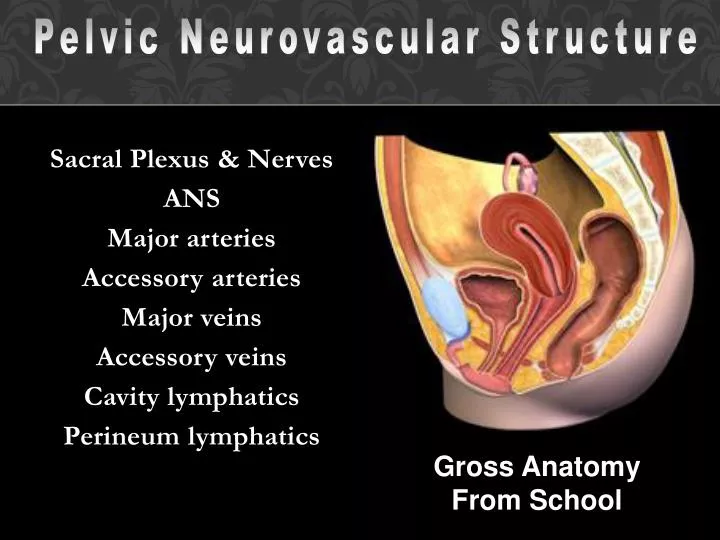
Pelvic Neurovascular Structure. Sacral Plexus & Nerves ANS Major arteries Accessory arteries Major veins Accessory veins Cavity lymphatics Perineum lymphatics. Gross Anatomy From School. A. Sacral Plexus.

E N D
Pelvic Neurovascular Structure • Sacral Plexus & Nerves • ANS • Major arteries • Accessory arteries • Major veins • Accessory veins • Cavity lymphatics • Perineum lymphatics Gross Anatomy From School
A. Sacral Plexus • Location: lies ant to& covered with pelvic fascia w/ nerves exiting in greater sciatic foramen
1. Sciatic n. – largest; major n. of LL a) comp: b) exits:
c) splits into: 1. Tibial n. –
2. Common fibular n. – superficial & deep fibular nerves
b) Exits: inf to piriformis m., crosses over lig, & re-enters in lesser sciatic f.
c) Enters Pudendal canal – medial border of neurovascular bundle
d) Pudendal n. in canal divides into: 1. Inf rectal n. – near entrance of pudendal canal a) motor: ext. anal sphincter & levator ani b) sensory: skin of anal triangle
2. Dorsal n of penis (clitoris) – descends in pudendal canal & in F = primary
a) Deep perineal n. – enters motor: muscles of deep & sup perineal pouches sensory: vestibule & inf. vagina
b) Superficial perineal n. – becomes posterior scrotal or labial n. - sensory
3. Other branches of sacral plexus: Name Spinal Seg Function Sup gluteal Inf gluteal N. to obturator internus N. to quadratusfemoris Post femoral cutaneous n Perforating cutaneous n. N. to piriformis N. to levatorani, coccygeus, ext anal sphincter
1. Sacral paravertebral trunk (2) – gangliaof the sympathetic trunk in pelvic cavity; a) courses over ala & descends medial to ant. sacral foramina b) Ganglion impar –
2. Sacral prevertebal trunk – carries sympathetic, parasymp, & visceral afferent fibers for pelvic organs &
3. Superior hypogastric plexus – distributes sacral prevertebal fibers to
4. Rt & lt hypogastric nerves – descend into pelvic cavity and connect with
6. Subsidiary plexuses – inn by inf. hypogastric plx: a) Rectal plexus b) Vesicle plexus
c) Uterovaginal (Prostatic) plexus – 1. Cavernous n. – from prostatic plexus erectile tissues of penis
D. Int. iliac Arteries 1. Def: major artery of the perineum 2. Origin: bifurcation of common iliac a. @ lumbosacral joint 3. bifurcates at pelvic brim into: ant. and post trunks
a) Anterior trunk – pelvic viscera, perineum, gluteal region, medial thigh, placenta b) Posterior trunk - post abdominal & pelvic walls + gluteal region of hip
4. Posterior Trunk (Prox dist): a) iliolumbar a. – ascends above pelvic brim along lumbosacral trunk: 1. iliac branch – serves iliac fossa 2. lumbar branch – lower post abdominal wall
b) Lateral sacral a (2) – ant. sacral foramina to serve sacrum & skin c) Sup. gluteal a. – greater sciatic notch, above piriformis to serve gluteal region
5. Anterior Trunk (prox dist): a) Umbilical a –adjacent to pelvic inlet ascends in ant. abdominal wall to umbilicus; one branch:
Superior vesical a.– serves distal ureter, superior surface of bladder, ductus deferens
b) Obturator a – below margin of pelvic brim, descends through obturator foramen w/ n. (above) & v. (below); serves medial thigh compartment
c) Inf. vesical (vaginal) a. – inf. bladder, ureter, seminal vesicle, & prostate (vagina), part of rectum)
d) Middle rectal a –serves rectum; anastomoses w/: 1. Superior rectal a – from inf. mesenteric 2. Inferior rectal a - from internal pudendal
e) Internal pudendal a. – main artery of perineum 1. greater sciatic foramen - inf to piriformis 2. re-enters in lesser sciatic f. thru pudendal canal
3. Branches of int. pudendal a.: a) Inf. rectal a. –ischioanalfossa middle rect. a. b) Perineal a. –pudendal canal sup. pouch w/ branches:
1. Post. scrotal (labial) a. – in superficial pouch 2. Dorsal artery of penis (clitoris) – through supplies superficial penis / clitoris & has two branches:
3. Deep artery of penis (clitoris) –terminal branch of perineal artery = serves the crus & corpora cavernosum
a) Artery of bulb of penis (vestibular bulb) – in deep pouch to serve corpus spongiusum b) Urethral a. – in deep pouch serves urethra
f) Additional anterior trunk branches in women:1. Uterine a –through broad ligament a) ascends lat. to uterine wall to uterine tubes anastomizes w/ ovarian a. b) descends lat to vaginal wall
g) Inferior gluteal a – terminal branch of anterior trunk exits in gr sciatic f., inf to piriformis
E. Non Internal-iliac Arteries 1. Median sacral a – @ bifurcation of abdominal aorta, descends in midline ant to sacrum, anastomizes w/ iliolumbar and lat. Sacral arteries
2. Ovarian a. – in suspensorylig, anast. With ant branch of int. iliac
3. Ext. pudendal a. – from femoral artery courses medially to serve anterior perineum skin
F. Internal Iliac Veins 1. Most veins follow arteries internal iliac v.