Download
1 / 31
370 likes | 650 Views
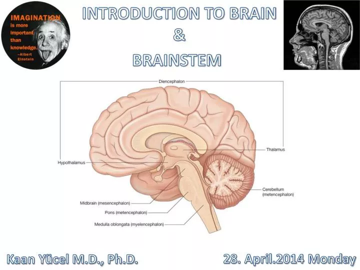
INTRODUCTION TO BRAIN & BRAINSTEM. 28. April.2014 Monday. Kaan Yücel M.D., Ph.D . The brain (encephalon) is divided into three major divisions. 1) Hindbrain ( Rhombencephalon ) I. Medulla oblongata II. Pons III. Cerebellum Pons and cerebellum are called as metencephalon .

E N D
INTRODUCTION TO BRAIN & BRAINSTEM • 28. April.2014 Monday • Kaan Yücel M.D., Ph.D.
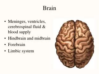
The brain (encephalon) is divided into three major divisions. 1) Hindbrain (Rhombencephalon) I. Medulla oblongata II. Pons III. Cerebellum Pons and cerebellum are called as metencephalon. 2) Midbrain (Mesencephalon) 3) Forebrain (Prosencephalon) I. Telencephalon (Cerebrum) II. Diencephalon (between brain)
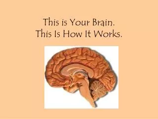
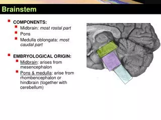
BRAINSTEM oldest part of the CNS. medulla oblongata, pons, and midbrain occupies the posterior cranial fossa of the skull. stalklike in shape and connects the narrow spinal cord with the expanded forebrain.
BRAINSTEM contains 10 cranial nerves, and most of the motor and sensory systems pass through this important region. a relatively small region (approximately 7 cm long) links the forebrain (i.e., cerebral cortex) and spinal cord and all messages going between the two areas must go through the brain stem.
Midbrain 2 cm in length connects the pons and cerebellum with the forebrain. cerebral hemispheres connected to brainstem by 2 large fiber tracts cerebral peduncles The dorsal aspect of the midbrain the tectum(L., roof] paired superior and inferior colluculi(singular, colliculus). corpora quadrigemina superior colliculicenters for visual reflexes inferior colliculilower auditory centers Tegmentum: Anteriorpart of midbrain BetweenCerebralaqueduct
Midbrain The midbrain comprises two lateral halves cerebral peduncles anterior part: crus cerebri substantianigra posterior part: tegmentum
Substantianigra • Largemotor nucleus between tegmentum & crus cerebri • Concernedwith muscle tone • Connectedto the cerebral cortex, spinal cord, hypothalamus, and basal nuclei.
Medulla [oblongata] • In the posterior cranial fossa, lying beneath the tentoriumcerebelli and above the foramen magnum. • Related anteriorly to the basal portion of the occipital bone and the upper part of the odontoid process of the axis and posteriorly to the cerebellum.
Medulla oblongata • Not only contains many cranial nerve nuclei that are concerned with vital functions (e.g., regulation of heart rate and respiration), but it also serves as a conduit for the passage of ascending and descending tracts connecting the spinal cord to the higher centers of the nervous system
BRAINSTEM Nuclei of 12 cranialnerves 10 of them in thebrainstem midbrain pons medulla Of the IV & III Of theother 4 VIII,VII,VI,V Of thelast 4 XII,XI,X, IX
I OlfactoryPurely sensoryTelencephalon Smelling II Optic SensoryRetinal ganglion cells Seeing III OculomotorMainly motorMidbrain Eye movements & pupillary reflex
IV TrochlearMotorMidbrain Intorts the eyeball. V TrigeminalBoth sensory and motorPons Receives sensation from the face and innervates the muscles of mastication. VI AbducensMainly motorPons Abducts the eye.
VII Facial Both sensory and motorPons Provides motor innervation to the muscles of facial expression. Receivesthe special sense of taste from the anterior 2/3 of the tongue and provides secretomotor innervation to the salivary glands (except parotid) and the lacrimal gland. VIII VestibulocochlearMostly sensoryPons Hearing and balance IX Glossopharyngeal Both sensory and motorMedulla Receives taste from the posterior 1/3 of the tongue, provides secretomotor innervation to the parotid gland, and provides motor innervation to the stylopharyngeus. Some sensation is also relayed to the brain from the palatine tonsils.
X VagusBoth sensory and motorMedulla Innervationto most laryngeal and pharyngeal muscles (except the stylopharyngeus, which is innervated by the glossopharyngeal). Parasympatheticfibers to nearly all thoracic and abdominal viscera till the proximal two-thirds of the transverse colon. Receives the special sense of taste from the epiglottis. A major function: controls muscles for voice and resonance and the soft palate.
XI Accessory (often separated into the cranial accessory and spinal accessory nerves) Medulla Mainly motor Cranial and Spinal Roots Controls the sternocleidomastoid and trapezius muscles, and overlaps with functions of the vagus nerve (CN X). Symptoms of damage: inability to shrug, weak head movement. XII Hypoglossal mainly motor Medulla Provides motor innervation to the muscles of the tongue (except for the palatoglossus, which is innervated by the vagus nerve) and other glossal muscles. Important for swallowing (bolus formation) and speech articulation.
Reticularformation • The reticular formation (L. reticulum, “little net”) consists of various distinct populations of cells embed in a network of cell processes occupying the central core of the brainstem. • The reticular formation and the olfactory and limbic systems are interrelated as a result of their participation in visceral functions and behavioral responses.
Reticularformation More than 100 nuclei scattered throughout the tegmentum of the midbrain, pons and medulla have been identified as being part of the brainstem reticular formation.
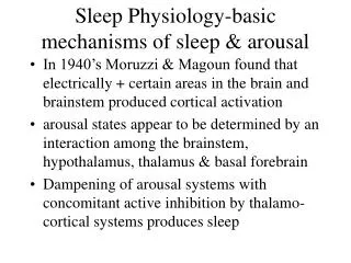
Reticularformation 1- The regulation of the level of consciousness, and ultimately cortical alertness 2- The control of somatic motor movements 3- The regulation of visceral motor or autonomic functions 4- The control of sensory information
Autonomic Nervous System SympatheticParasympathetic Anatomicaldifferences, differences in the neurotransmitters, differences in the physiologic effects The autonomic nervous system and the endocrine system control the internal environment of the body. The various activities of the autonomic and endocrine systems are integrated within the hypothalamus.
Autonomic Nervous System Sympatheticpart prepares and mobilizes the body in an emergency, when there is sudden severe exercise, fear, or rage. Parasympatheticpart aims at conserving and storing energy, in the promotion of digestion and the absorption of food by increasing the secretions of the glands of the gastrointestinal tract and stimulating peristalsis.
Autonomic Nervous System • Parasympatheticsystem • Brainstemand sacral segments of the spinal cord • Edinger-Westfall nucleus • midbrain • mediates the diameter of the pupil in response to light • Superior and inferior salivatory nuclei • pons &medulla • mediatie salivary secretion and the production of tears) • Dorsal motor nucleus of the vagus nerve • Medulla • controls the motor responses of the heart, lungs, and gut • (e.g., slowing of the heart rate and constriction of the bronchioles). .
Cerebralaqueduct (aqueduct of Slyvius) • A narrowchannelconnectingthirdandfourthventricles • Linedwith ependyma • Surroundedby a layer of gray matter:central gray • Directionof flow of CSF 3rd ventricle 4 thventricle • No choroid plexus